Why it’s always good to (also) scan the chest when performing an abdominal echo
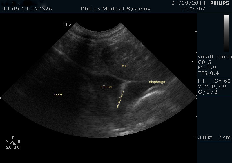
So, this lovely 11-month-old French Bulldog presented with a 4 day history of innappetence, vomiting and retching. Her abdominal scan was pretty unremarkable but from a sub-xiphisternal window a pleural effusion was visible.
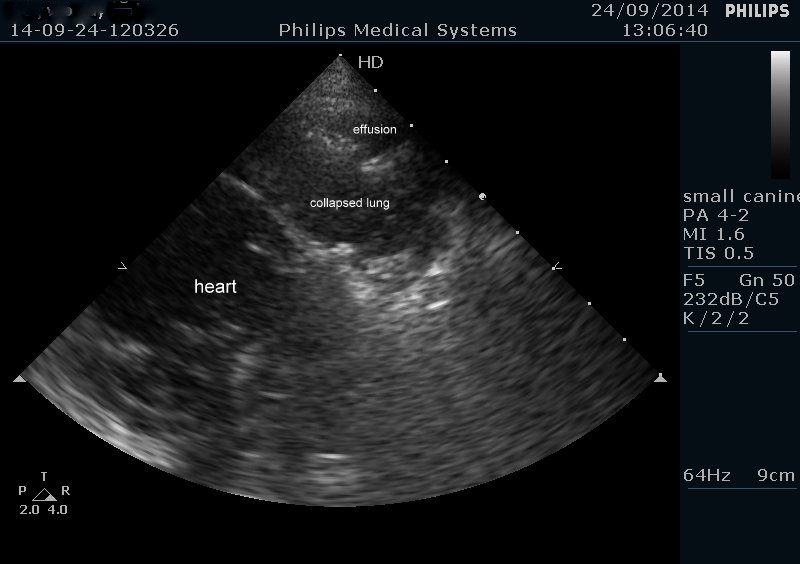
The caudal mediastinum is visible due to the bilateral nature of the pleural effusion. However, on further investigation the left hemithorax was found to be more severely affected and, furthermore, there was isolated collapse of the left cranial lobe.
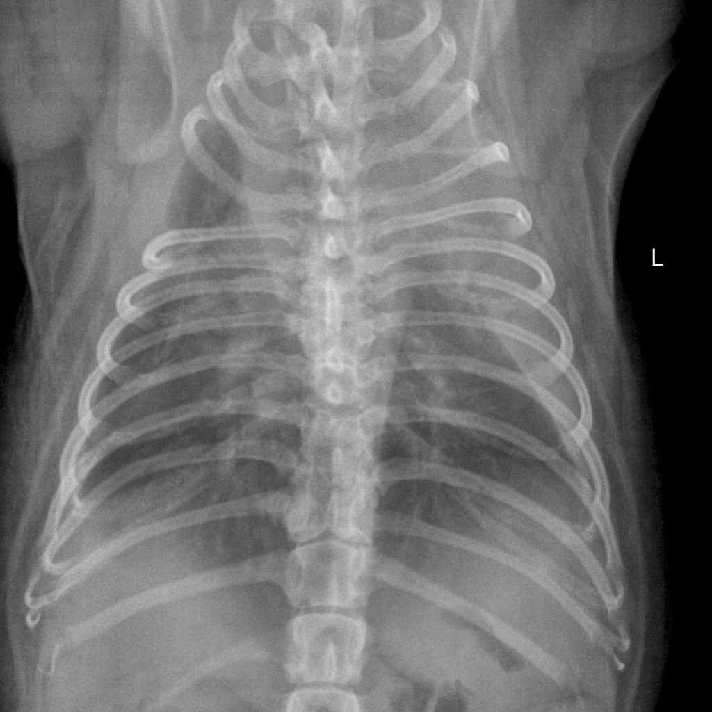
You can tell it’s likely lung (apart from the fact that it’s in the right place!) because there are just a few hyperechoic gas-filled spaces near the periphery. So we tapped the effusion and it was bloody -in fact, cytologically, a sterile haemorrhagic exudate. To x-ray…….
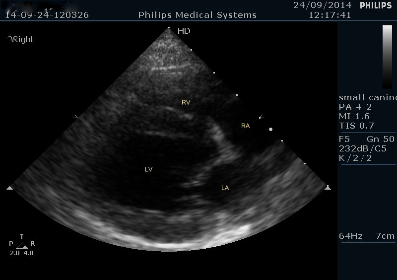
Hmmm….a bilateral effusion and confirming collapse of the left cranial lobe. With a haemorrhagic effusion surely that’s most likely a lung lobe torsion -the left cranial is the predisposed lobe in small breed dogs (whereas big, deep-chested dogs twist their right middle lobes). How about the possibility of pulmonary thromboembolism? Well there was a bit of tricuspid regurgitation but it wasn’t high-velocity-enough to fit with pulmonary hypertension as usually seen with severe PTE. And the cardiac chambers are in normal proportion -where we would expect right heart dilation with acute PTE.
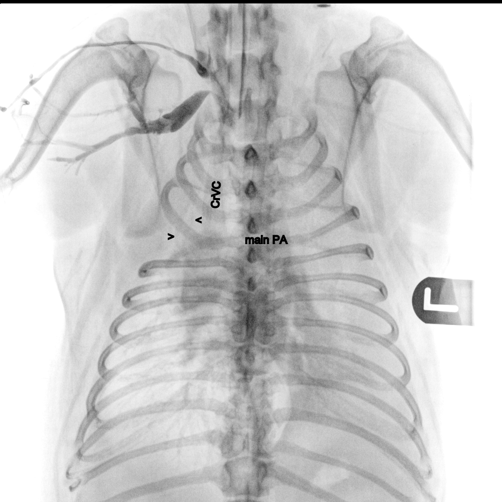
Since it was easy to do, we also ran a little non-selective angiogram (omnipaque bolus into right cephalic vein).
Right of the cranial vena cava the arrowheads indicate the right cranial lobe bronchi. And, sure enough, there appears to be no perfusion of the left cranial lobe.
At surgery there was indeed a flipped-over left cranial lung lobe -heroically removed by Matt and Nicole at Yorkshire Vets, Thornbury. She’s doing well at the time of writing.


