‘Urinary cyst’ in a dog: why you should measure creatinine in prostatic cyst fluid
Prostatic cysts are a common finding -particularly in entire male dogs. These are aetiologically distinct from para-prostatic cysts which are thought to form within remnants of the mullerian ducts adjacent to the prostate. Intra-parenchymal microcysts are almost always present when hyperplasia is pronounced. Larger fluid-filled spaces are often termed ‘retention cysts’: these generally contain prostatic secretions. However, a proportion also fill with urine -a result of spontaneous urethral fistulation: so-called ‘urinary cysts’.
A nice review has been written by Bokemeyer et al.
http://onlinelibrary.wiley.com/doi/10.1111/j.1748-5827.2011.01039.x/abstract
These authors advocate the prospective measurement of creatinine in prostatic cyst fluid in order to identify pre-surgery those cases with fistulation.
16/87 (18%) dogs with prostatic cavitary lesions in their series turned out to have ‘urinary cysts’: using cyst fluid creatinine at least twice plasma creatinine as the discriminating criterion. Radiographic contrast studies can also be used to demonstrate communication between urethra and cyst cavity.
Identifying urine-filling is useful because these often fail to resolve after castration and percutaneous drainage. Although, in this series, some dogs were lost to follow up there were only two cases where non-surgical cyst management was known to have been successful. In the remainder, either surgery was required because of recurrent filling or the problem remained unresolved due to owner unwillingness for intervention.
The surgical procedure usually involved opening the cyst and identifying the fistula by direct visualisation of an exposed urethral catheter or by use of urethrally-introduced methylene blue dye. Fistulae were sutured closed and the cysts were subsequently omentalised.
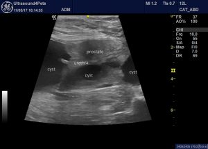
This is the prostate of just such a case seen recently by ourselves:

Long-axis view of the prostate
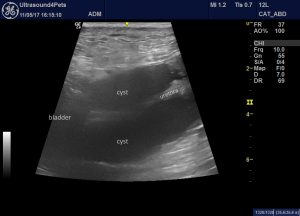
The cyst is partly within the prostate but also extends both cranially and caudally. Most significantly, the cyst cavity is directly in apposition to the urethra. In fact it completely encircles the exposed urethra at one point:

long axis view of the urethra within the cyst cavity caudal to the bladder neck
Communication between the cyst and the urethra was demonstrated with positive contrast urethrography.
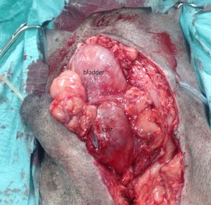
At surgery the cyst is apparent encircling the proximal urethra:

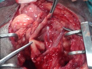
And when opened the defect in the urethral wall was readily located with a urethral catheter:

After surgery a urethral catheter is usually left in place for a few days to relieve pressure on the closure of the urethra.
Full details of the surgery can be found here from our excellent scalpel-wielding colleagues at West Midlands Referrals:


