Ultrasound lung rockets
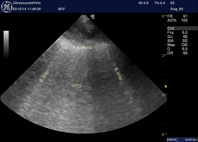
Hot topic of the moment in veterinary ultrasonography this is. Images below from an old dog with acute severe malaise (to the extent of recumbency) but few specific signs. Haematology and biochemistry relatively unremarkable. Abdominal ultrasonography similarly so. However, ultrasonographic survey of the chest revealed a site on the lower left chest wall with abnormal findings.
Those bright white lines arising at the pleura and radiating away from the probe are known as B lines, or (more dramatically!) ultrasound lung rockets, and are indicative of ‘wet lung’ -i.e. conditions such as pulmonary oedema, pneumonia or contusions. In a moving image they glide from side to side with respiration as the lung moves against the chest wall. The pleura is also thickened and slightly irregular in this dog and there may be a very small pleural effusion.
Recent papers show that in standing or sternally-recumbent dogs B lines are very few and far between. More than one or two in a selection of intercostal sites indicates likely increased lung fluid content. It’s a very useful indicator of lung issues and a great way to monitor fluid therapy (where the appearance of B lines indicates overhydration) or treatment of pulmonary oedema (where their disappearance indicates success).
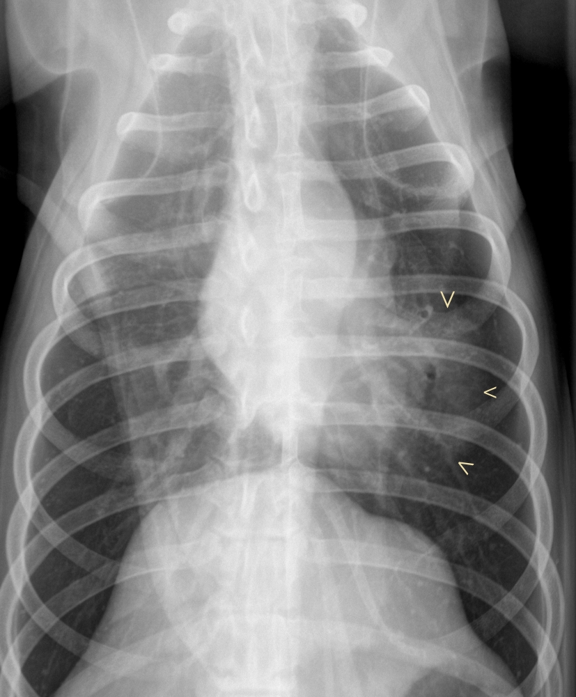
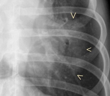
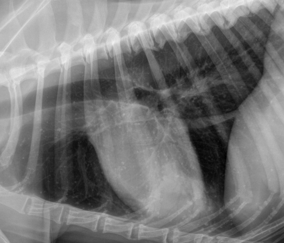
Localised areas with B lines such as this are consistent with lung diseases such as pneumonia or contusions. In this case the history suggests that pneumonia may be the most likely cause. Chest radiographs were not dramatic -we could easily have overlooked the abnormalities if we hadn’t had the ultrasonography first.
However, there clearly is a consolidated peribronchial area in the ventral left lung.
With a provisional diagnosis of possible pneumonia we started intravenous antiobiotics and the patient responded promptly within 24 hours. I’m sure with hindsight that we have under-diagnosed pneumonia in dogs in the past but maybe thoracic ultrasonography will help in the future.


