Ultrasound-guided lung biopsy
One more on the topic of ultrasonography of lung lesions.
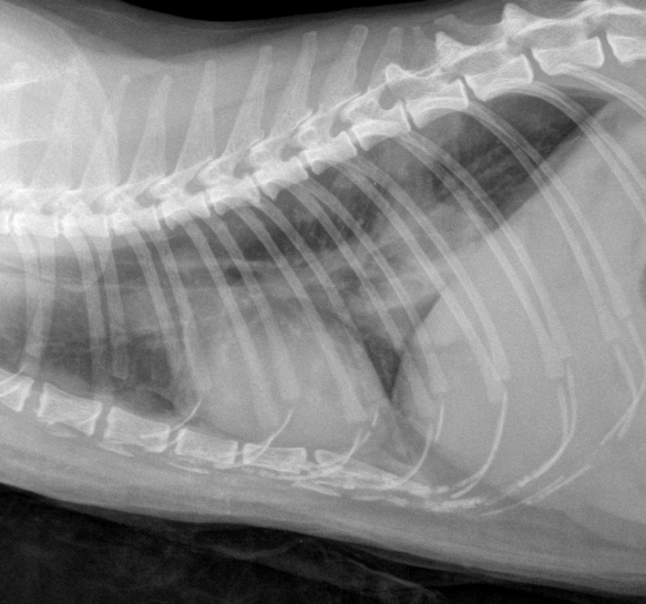
This is a 9 year-old cat with a cough. Nothing outwardly dramatic. Admittedly thoracic radiographs look a little concerning:
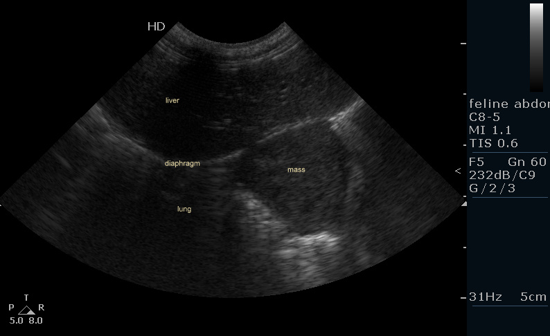
…but we’ve all seen lots of cats with asthma that look something like that. On the other hand, take a look at the ultrasonogram looking forward through the diaphragm.
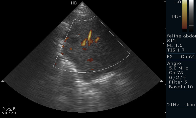
That doesn’t look so much like asthma any more. And with power Doppler……
Not only is there a solid soft tissue mass; it’s a very vascular mass -not an abscess or collapsed lung by the looks of it.
[wpvideo oBYzXxaK]
Ultrasound-guided lung biopsy like this is best done under general anaesthesia. Hold the lungs inflated and immobile by applying positive pressure on the bag of the T-piece: this avoids the needle ripping through lung tissue with respiratory movements. With this kind of solid mass the risk of iatrogenic pneumothorax is minimal.
Histology came back as an adenocarcinoma: probably a primary lung tumour. The causal lobes are the predisposed site. in dogs they are well worth removing with lung lobectomy as survival times > 1 year are not uncommon. In cats it’s not so good.


