The significance of bright bits in the pancreas. Ultrasound elastography (USE) in pancreatitis
Ultrasound elastography, or elastosonography, is increasingly available on high-end ultrasound machines;. At the most basic level USE gives an indication of tissue stiffness. This may be either in response to mechanical pressure (strain imaging) or to ultrasound-generated compression (shear wave elastography). Mechanical pressure in strain imaging is exerted externally (manually) or by internal changes due to respiratory or cardiovascular movements. In either case, changes in echo signal indicative of varying stiffness are colour-coded in the screen image.
Theranostics. 2017; 7(5): 1303–1329.
Ultrasound Elastography: Review of Techniques and Clinical Applications
Rosa M.S. Sigrist,1 Joy Liau,1 Ahmed El Kaffas,1 Maria Cristina Chammas,2 and Juergen K. Willmann
https://www.ncbi.nlm.nih.gov/pmc/articles/PMC5399595/
An excellent summary of USE in veterinary medicine is this presentation by Cibele Figueira Carvalho from IVUSS 2014:
http://www.ivuss.org/wp-content/uploads/resources/SalzburgNotes/Elastosonographylecture.pdf
Strain imaging is the prototype USE modality. Shear wave technology represents a further advancement but remains significantly more expensive.
In human medicine, USE has been useful in assessing liver and kidney fibrosis and in differentiating malignant vs benign lesions in liver (sensitivity and specificity of 97% and 66% respectively in one study), breast and thyroid tissue amongst others.
This preamble brings us to images generously contributed from Canada by Bob Hylands in response to earlier discussion in these blog posts of the significance of hyperechoic intraparenchymal ‘flares’ or ‘marbling’ as a possible marker of pancreatitis.
Some more examples here:

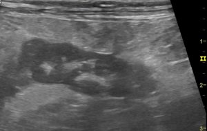
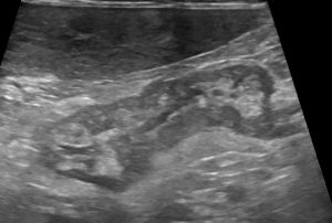
The tail of the right lobe of a dog with ‘acute abdomen’. This is what I would call classic ‘marbling’: angular, intensely hyperechoic foci in a hypoechoic pancreas. I would interpret this as presumed acute pancreatitis -perhaps with saponified fat?

A canine left pancreas: this looks slightly less acute. The hyperechoic foci are rounded. Maybe this is fibrosis?
OK, so these are Bob’s images of feline pancreatitis with elastography:

Feline pancreatitis with elastography: the hyperechoic ‘marbling’ is revealed to be softer (coded red on elastography) than the surrounding parenchyma (images courtesy of Bob Hylands)
Those hyperechoic foci are shown to be softer/more elastic than the surrounding hypoechoic pancreas. That suggests that they may be saponified fat rather than fibrosis…at least in this individual cat.
Hyperechoic abdominal (mesenteric) fat may result from a variety of pathological processes and is especially difficult to assess in the presence of ascites. Intrapancreatic fat saponification might be a more specific marker of pancreatitis.
Any opinions welcome.


