Ultrasonography of the canine and feline caecum (or cecum)
There’s very little in the published literature about scanning the caecum in small animals -which is surprising since it is often encountered during abdominal examinations.
Although dogs often have more gas in their colons and caeca, which sometimes makes it more difficult to make out, the canine caecum is also quite an elongated structure (usually about 5cm) which runs across the middle of the abdomen and may be seen from several views.
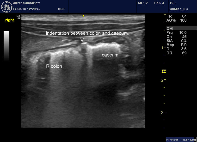
From the right side of the abdomen the ileum, ileo-caeco-colic junction, right colon and caecum lie deep to the duodenum. It’s almost always fairly simple to find right colon (it’s the big gas-filled thing which makes it hard to see past) and then it’s a matter of exploring both cranially and caudally until the entry of the ileum is found. Exploring around the ICCJ will reveal the caecum in one plane or another. Sometimes you can just see a big gas filled right colon and then an indentation in the surface at the junction with the gas-filled caecum -as in this view (this is a cat):
From a right, dorso-lateral viewpoint, looking at the caudal vena cava, the caecum often lies immediately ventral to the cava.
From the left, the tip of the caecum quite often appears deep to the spleen in the same area as the left pancreas.
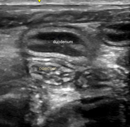
When relatively empty it is of similar diameter to small intestine but with the wall thickness of large intestine. The inner contour is corrugated. This is a longitudinal plane image from the right ventral abdomen with the caecum seen in short-axis deep to the duodenum:
This video from the right flank of a dog in longitudinal plane shows the caudal pole of the right kidney, the descending duodenum superficially and the caecum (deep to the duodenum and caudal to the kidney) which communicates with the right colon (which in this case, happily, contains faeces but minimal gas) as the probe is fanned laterally..
[wpvideo RkYRYRqI]
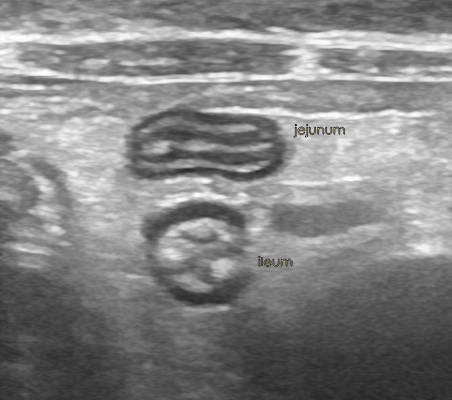
In cats, the ICCJ is easier to find because 1) the abdomen is smaller 2) there is often less gas in the large intestine and 3) the ileum has a very distinctive appearance with a thick, crinkly, hyperechoic submucosa.
Once the ileum is found then the ICCj and the caecum aren’t far away. It’s a matter of exploring in several planes to find the 3 pieces of gut (ileum, right colon, caecum) which are joined at the ICCJ.
OK, so from a clinically-relevant point of view the first thing is that it’s good to know what a normal caecum looks like so that you don’t misinterpret it as a loop of diseased small intestine.
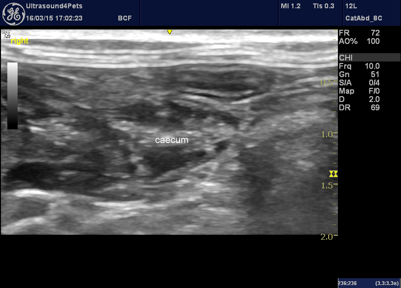
Secondly, pathological things can happen in caeca. This is a cat with a typhlitis (inflamed caecum) possibly due to Tritrichomoniasis :
The mucosa is very thickened.
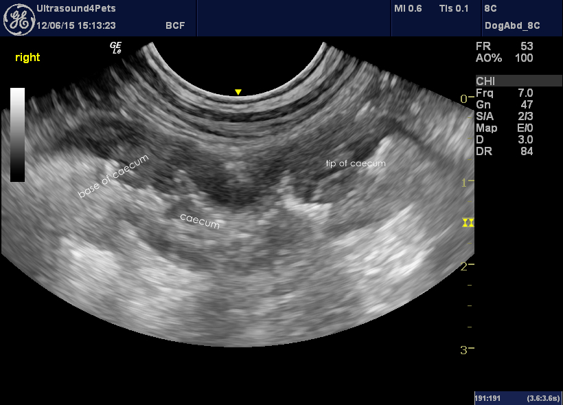
And this is an interesting dog who presented with abdominal pain and a history of chronic anal furunculosis.
That’s a right sided, longitudinal plane view which captures the entire caecum from its base cranially to tip caudally. The wall is massively and irregularly thickened up to 6mm. Although we don’t have a definitive diagnosis in this case there does appear to be an association between anal furunuclosis and colitis in some cases (e.g. Jamieson et al JSAP Mar 2002) so it is tempting to speculate as to a common cause.




I want to know what typhlitis mean in dogs?
inflamed caecum