Ultrasonography of canine bladder wall masses
Now, this is a common problem. Dogs with dysuria and/or haematuria. You perform a full abdominal ultrasonogram and, behold! – an apparent soft tissue mass in the bladder.
So, what is it? An FNA is generally regarded as being inadvisable since transitional cell carcinomas may seed along the needle line. Cystoscopy is certainly an option but it can be difficult to get a diagnostic sample with the small gauge biopsy forceps which will fit through a cystoscope and it can be hard to get a biopsy from lesions in the bladder neck . As an occasional cystoscoper I find it a technically challenging procedure. Urine cytology may help: but this is very dependent on your cytologist’s willingness to make a call on the distinction between reactive and neoplastic urothelial cells. Bladder tumour antigen testing may help: but blood contamination leads to lower specificity (although a negative test is a valuable indicator that TCC is less likely since the test is @90% sensitive).
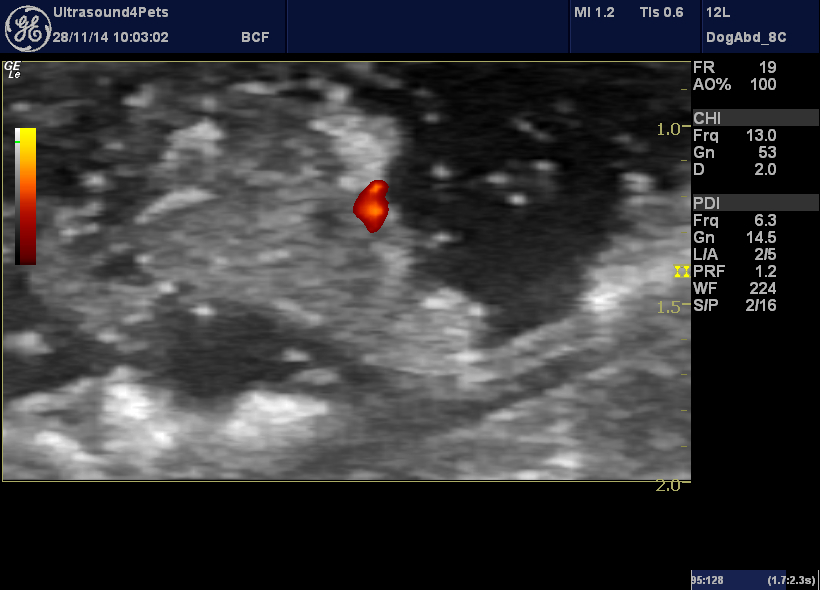
My first step is always to have a closer look at the ultrasound images. There is more information here. The first step is to establish that the apparent mass really is a mass and not a clot. Blood clots can appear deceptively mass-like. It’s embarassing when the ‘tumour’ has vanished 2 weeks later. If the patient can be persuaded to lie still enough then power Doppler will tell you if the mass is vascularised -in which case it’s not a clot!
That red splodge at the caudal margin (they must be consistently present to rule out movement artefact) is pulsatile and represents an arterial vessel.
There is a recent and excellent review article looking at ultrasonographic features of transitional cell carcinomas in dogs:
Ultrasonographic findings related to prognosis in canine transitional cell carcinoma.
These authors found that important indicators of negative prognosis (due to neoplasia) were trigone site, heterogeneous mass appearance and invasion of the bladder wall.
So, our mass is certainly very heterogeneous. And there is also wall invasion:
Normal dorsal bladder wall can be seen to the right of the tumour attachment. The normal layering of hyperechoic lumen/mucosa interface, hypoechoic mucosa, hyperechoic submucosa, hypoechoic muscularis and hyperechoic serosa is the same as in gut wall. At the base of the mass there is clear disruption and invasion into the muscularis -the hallmark of bladder wall neoplasia. Polyps are confined to the mucosa.


If the gas in the adjacent descending colon makes the cystic calculi difficult to detect, the patient can be scanned while standing, using the benefits of gravity to mobilize the calculi away from the dorsal urinary bladder wall.
Thanks Kioin, that’s really true: good idea to scan them in multiple positions to see if things in the bladder are mobile.