Ultrasonographic findings in feline exocrine pancreatic insufficiency (EPI)
This is just a snippet but maybe worth including because I’m not aware of any published images of the pancreas in cats with EPI.
For example, there is no mention of imaging in this recent case series:
https://www.ncbi.nlm.nih.gov/pmc/articles/PMC5115185/
J Vet Intern Med. 2016 Nov-Dec; 30(6): 1790–1797.
Feline Exocrine Pancreatic Insufficiency: A Retrospective Study of 150 Cases
P.G. Xenoulis,corresponding author 1 D.L. Zoran, 2 G.T. Fosgate, 3 J.S. Suchodolski and J.M. Steiner
The present case being a 10 y.o. British Shorthair with severe weight loss, good appetite and steatorrhoea.

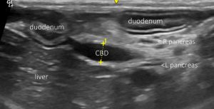
Longitudinal view of the porta hepatis from the right side: the common bile duct is borderline dilated (3.5mm) but not obviously obstructed by cholelith or mass. The pancreas was very difficult to find convincingly in this standard view. However, the ducts of the right and left lobe (labelled) could be followed down to the duodenal papilla.
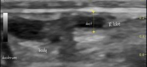
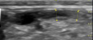
With more hunting around the base of the right lobe was located: this being the only area with any appreciable volume of pancreatic parenchyma:

longitudinal view of the right pancreas from the right side

The left lobe could not be identified from the left flank. There was no appreciable hyperechogenicity of peripancreatic fat.
Normal diameter of the right lobe according to various texts should be 3-6mm.
Serum TLI was markedly subnormal.


