Ultrasonographic diagnosis of meniscal tears in dogs
This is still an evolving area but very promising thus far. It’s relatively simple to scan the medial and lateral parts of the meniscii in dogs. A linear probe with frequency >10Hz will do the job nicely. A full ultrasonographic examination of the canine stifle includes views from cranial, cranio-lateral, cranio-medial, medial, lateral, caudo-medial and caudo-lateral. A critical assessment should be made of the patellar and collateral ligaments, presence/absence of joint effusion, joint capsule changes, meniscii, articular cartilage and bone surfaces. It is not easy to see the cruciate ligaments.
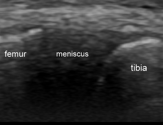
The healthy meniscus, viewed from caudo-medial or caudo-lateral appears as a neat, homogeneous, triangle of hyperechoic tissue wedged between the tibia and femoral condyle.
Figure: a healthy medial meniscus as seen from a caudo-medial approach
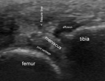
Meniscal tears and/or degeneration are readily apparent in many dogs with stifle problems. Typically there is joint effusion. The meniscus often bulges or slips medially (medial meniscus) or laterally (lateral meniscus) and the outer margin often appears ‘frayed’. The normal homogeneous echotexture is usually disrupted with hypoechoic zones and, if there is a tear, fissure lines are visible dividing the meniscus into discrete ‘slabs’.
Figure : damaged medial meniscus associated with cranial cruciate rupture -as seen from a caudo-medial approach. The meniscus is prolapsed medially. Its echotexture is quite heterogeneous and the free border is frayed.
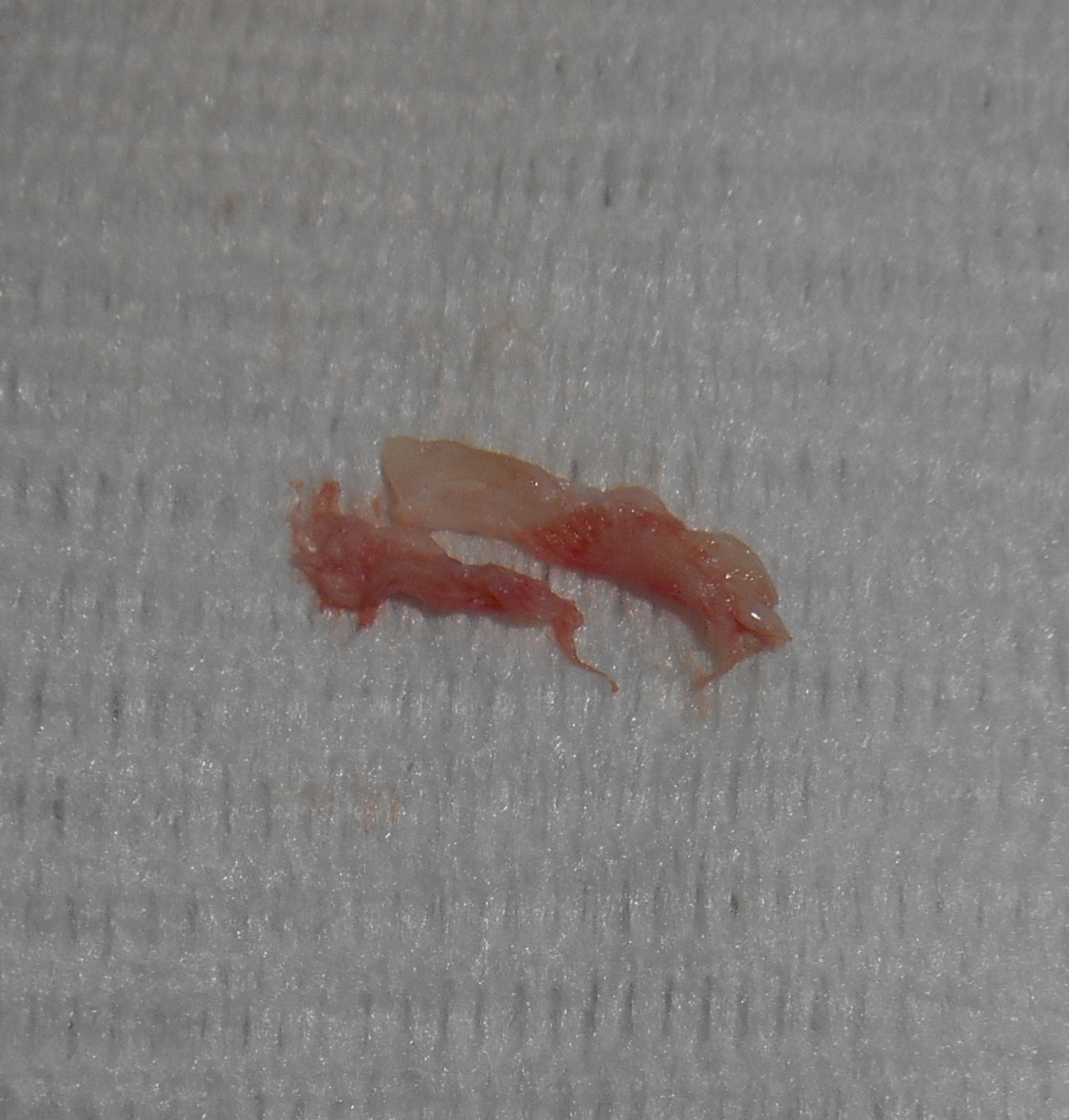
Figure: this is part of the medial meniscus (after partial meniscectomy) seen above. At arthrotomy the caudal part was torn with a well developed flap.
It is still early days but thus far, all cases which we have suspected ultrasonographically have been borne out at surgery. The best published paper (Arnault et al. Veterinary Comparative Orthopaedics & Trauma 6/2009) found sensitivity and specificity of 82% and 93% respectively. This is especially useful now that it is not necessarily seen as obligatory to perform arthrotomy during surgery for cruciate rupture. It’s also very helpful when faced with a possible ‘late’ meniscal injury post-surgery.


