Spontaneous pneumothorax in a cat
A previously-healthy, three-year-old, indoor DSH was presented with a 2 day history of cough and deteriorating respiratory distress.
These are the thoracic radiographs:
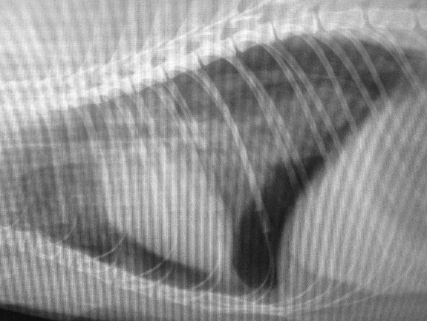
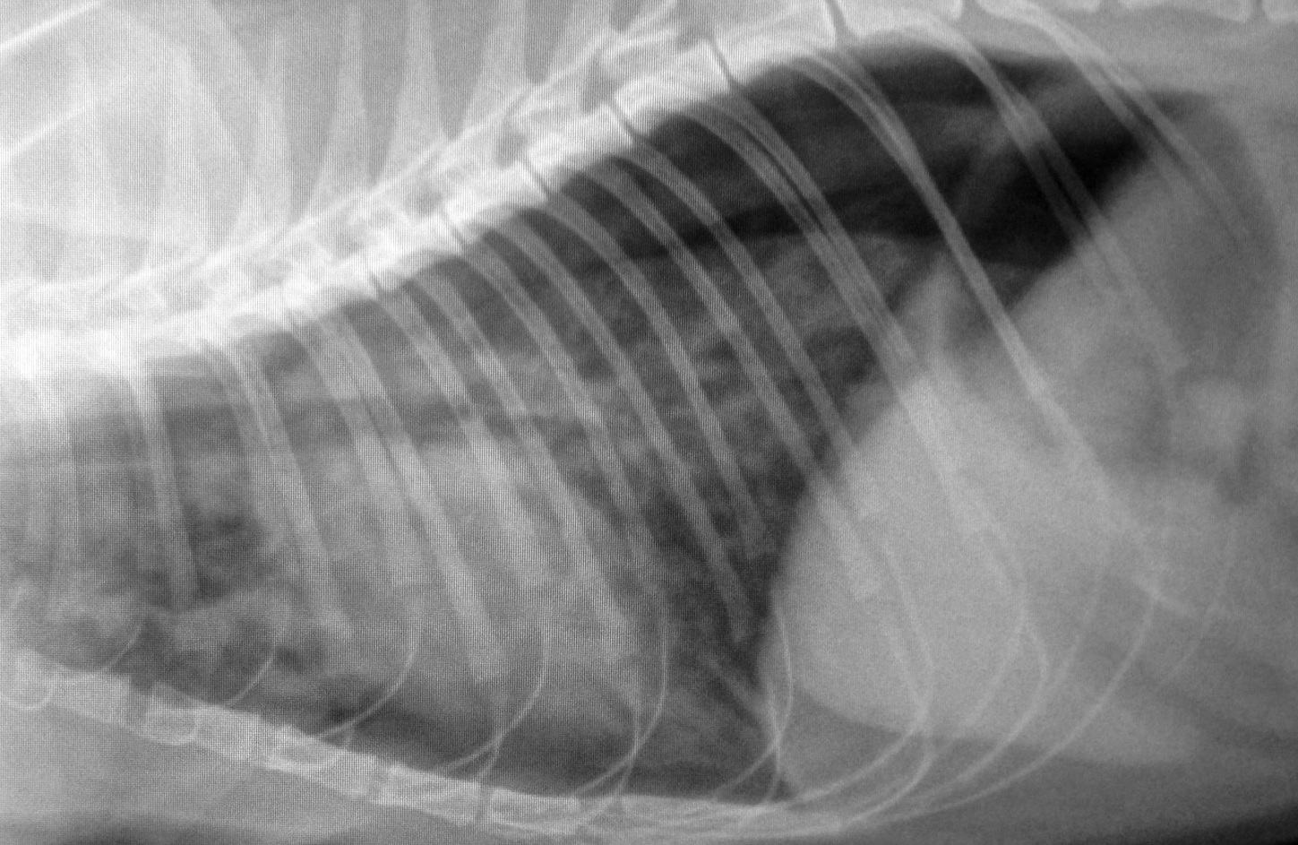
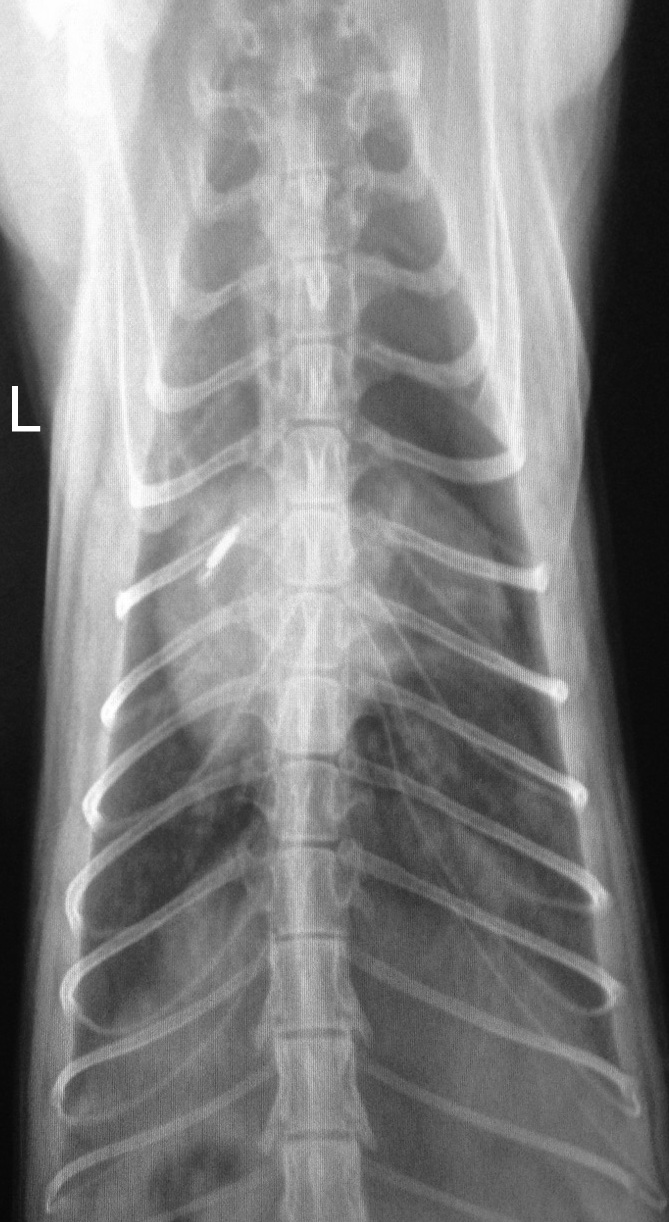
Looks pretty suspicious for a right sided pneumothorax. This is cat who has definitely no contact with motor vehicles. But….spontaneous pneumothorax is certainly a possibility:
http://www.ncbi.nlm.nih.gov/pubmed/24697868
http://www.ncbi.nlm.nih.gov/pubmed/22344603
It’s not common and, as these authors reported, most affected cats have lung disease.
On ultasonography:

R long axis four-chamber view
Left atrial diameter is nothing untoward, there’s no septal bowing or any evidence of thrombi. The appearance of the left ventricular wall in this frame is misleading -there was no thickening.
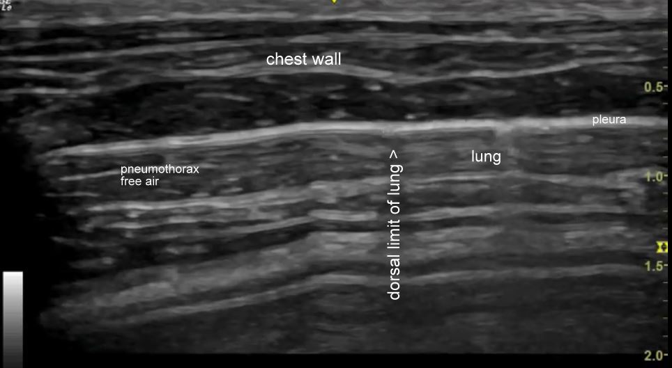
The thorax is very interesting though. This is a transverse plane view along the length of an intercostal space on the right side:
After a few seconds of wandering around the clip settles down to a view with a ‘glide sign’ on the right (ventral) where the partially collapsed lung is sliding against the chest wall. To the left (dorsal) there is no glide sign as there is just air below the parietal pleura.
In a still image frozen from the video clip:
With M mode, the dorsal part of the chest with pneumothorax looks like this:

…and the ventral part like this:

That’s a ‘sandy beach’ sign!
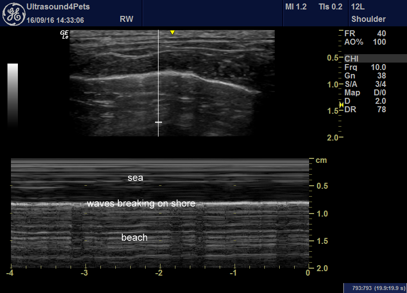
Pneumothorax is associated with loss of ‘sand’ texture. This requires a bit of practice but can be a very useful, quick, no restraint required technique for diagnosis of pneumothorax.
This cat’s lungs look abnormal on ultrasonography. That may be partially because they are somewhat collapsed but it also fits with the premise that there may well be lung pathology. My guess is that this cat has asthma -it comes at the end of a still, hot week with poor air quality and the patient is otherwise very healthy.


