Spleno-azygous shunt in a dog
This is just an excuse for some nice images. The patient is a 3 year old Lhasa apso with signs and bile acids consistent with a portosystemic shunt.
Longitudinal plane images from the right side behind the last rib looking at the area of the great vessels and porta hepatis.
The vena cava is clearly seen ventrally and to the right (nearer top of picture) and, even in a black and white image, flow looks fairly laminar within it.
Dorsally and to the left is the pulsing aorta.
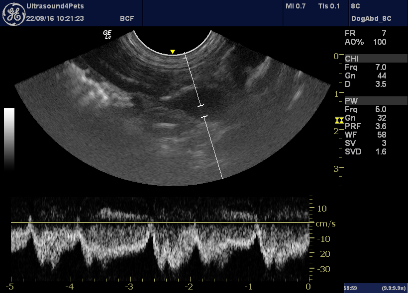
Between them is an abnormal, large vessel with cranially-directed flow. Where is this coming from???
OK so that’s a bit complex. This series of frozen frames has annotations:
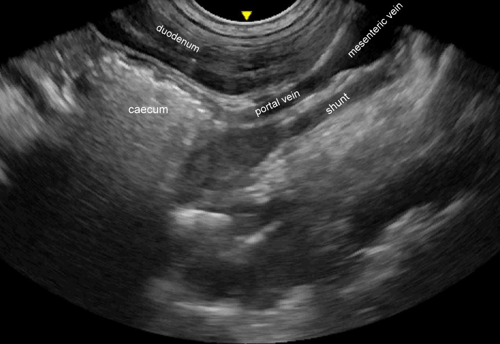
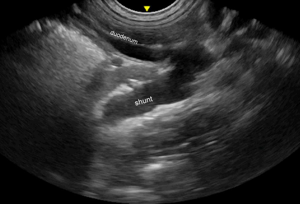
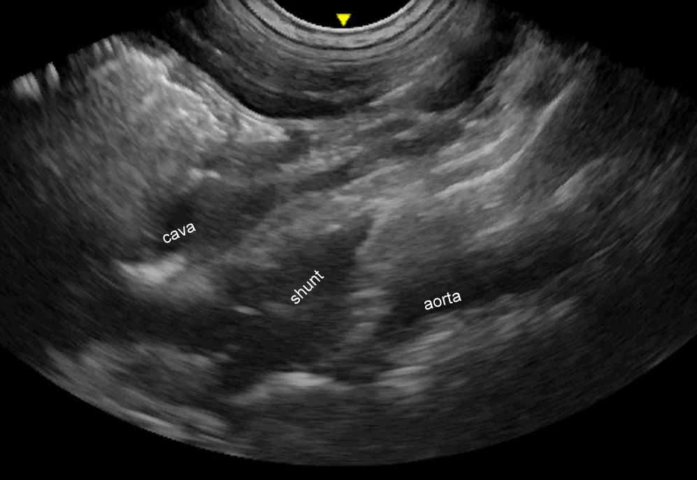
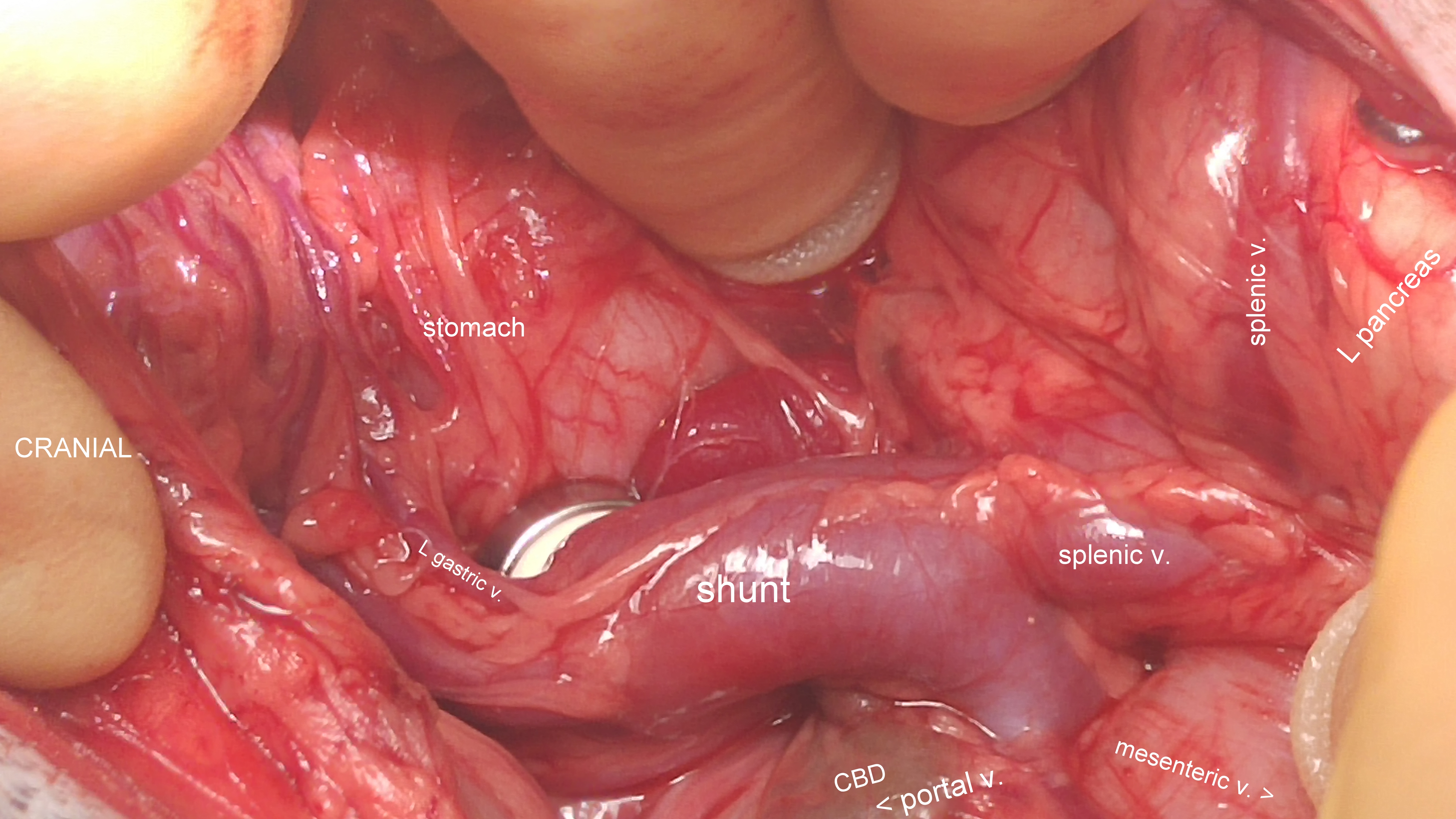
The shunt originates from the base of the splenic vein where the left gastric vein forms the first part of the shunt pathway. It then courses dorsally towards the aorta and joins the azygous vein before crossing the diaphragm.
Flow through the shunt is typically fluctuant -velocity changes with respiratory cycle. Whereas flow in the portal venous system and its tributaries is usually even (unless the patient is panting or hyperpnoeic).
Presentation relatively late, in adulthood, is a feature of azygous and phrenic shunts.
This is the offending vessel at surgery with an ameroid constrictor applied:
