Splenic vein congestion in GDV
This is a graphic illustration of what happens to the portal circulation in GDV cases. The torsion of the mesenteric attachments and pressure from the inflating stomach cause occlusion of the main portal vein. This is an important cause of abrupt reduction in venous return to the heart and subsequent catastrophic fall in cardiac output.
Portal vein occlusion causes pressure to back up into its tributaries -including the splenic vein. In this video the distended splenic vein contains static blood. This stasis is contributing to a degree of spontaneous echocontrast which means that the direction of blood flow can be seen without Doppler. On inspiration the blood ebbs backwards a bit (hepatofugal) and then flows forwards (hepatopetal) on expiration.
[wpvideo tbQ4tIYI]
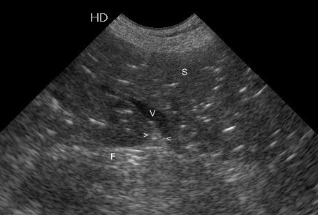
This poor dog is very acutely affected. After a few hours, where there is splenic torsion, the splenic pulp takes on a ‘starry sky’ appearance with hyperechoic microbubbles on a hypoechoic parenchyma. The image below is from a different case.
Splenic torsion often also creates hyperechoic triangles of mesenteric fat adjacent to the exit point of splenic veins (indicated by arrowheads in above image). This is thought to be due to swelling of the surrounding splenic parenchyma (….it can also be seen in other causes of marked splenomegaly). In the video clip there is a strong suspicion of this feature although the ‘starry sky’ effect has yet to develop.


