Splenic torsion….and the catchily-named hilar perivenous hyperechoic triangle sign
*Advanced warning -gory surgical photo involved here*
This Great Dane presented with acute, non-tympanic abdominal distention and malaise.
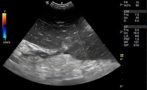
On sonography the spleen exhibits the classic ‘starry sky’ parenchymal pattern which is typical although not specific for torsion:

There is no detectable flow in the splenic vessels with Doppler.
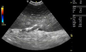
At the point where the splenic veins cross the capsule there are hyperechoic triangles of mesosplenic fat (arrowheads):

This is an ultrasonographic sign in need of a good name!
The hilar perivenous hyperechoic triangle as a sign of acute splenic torsion in dogs.
Again, this is typical but not completely specific. Any pathology which results in splenic distention (e.g. extensive acute infarction) could theoretically cause this effect -but torsion is the commonest in dogs. My understanding is that the splenic capsule bulges between veins but remains fixed by its attachment at the point of vein exit -creating an indentation filled by fat.
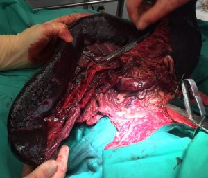
And at surgery…


