Sonography of canine interstitial lung disease: some examples
Firstly, a young CKCS with PCR +ve Pneumocystis pneumonia: this dog had a history of masticatory muscle myositis managed successfully with chronic steroid therapy until he recently developed severely increased respiratory effort.
There are widespread lines perpendicular to the pleurae. As discussed in previous posts, the appearance of abnormal lung lines on ultrasound is a whole can of worms; being very dependent on probe choice and settings. There’s pretty much a continuum in the appearance of vertical lung lines: from ‘classic’ B lines with bright ‘laser like’ appearance running down to the bottom of the screen; through broader C lines all the way to short, indistinct lines compatible with i lines as described in the literature.
In this dog with Pneumocystis there are clearly small subpleural consolidations and thickening/irregularity of the pleural line.
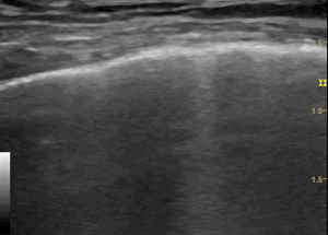
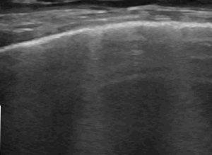
The thick, vertical, hyperechoic stripes (sometimes clearly associated with pleural/subpleural lesions) are what I would call ‘C lines’. These are generally considered highly indicative of pneumonia/pneumonitis in human medicine.
Secondly, a dog with apparent steroid-responsive interstitial pneumonitis. She presented with a week long history of anorexia, antibiotic-unresponsive fever and occasional retching/gagging. Sonography revealed no convincing abnormalities of abdomen, heart or neck. However, in two areas she exhibited marked lung changes again most compatible with C lines.
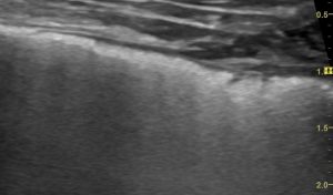
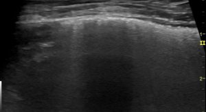
This dog responded promptly and completely to steroid therapy with no relapse. Obviously, it’s hard to definitively confirm a diagnosis of immune-mediated pneumonitis (sometimes referred to as ‘cryptogenic organising pneumonia’) in a dog which recovers.
Steroid-responsive idiopathic interstitial lung disease in two dogs
Liza Koster, Robert Kirberger
It seems to us that hyporexia and fever (rather than respiratory issues) tend to be dominant presenting signs in this condition.
There’s a really nice paper from human medicine looking at M mode in distinguishing cardiogenic pulmonary edema (CPE) from non-cardiogenic alveolar interstitial syndrome (NCAIS).
Chest 2018 Mar;153(3):689-696
The Use of M-Mode Ultrasonography to Differentiate the Causes of B Lines
Anup K Singh, Paul H Mayo, Seth Koenig, Aranabh Talwar, Mangala Narasimhan
https://journal.chestnet.org/article/S0012-3692(17)32961-6/pdf
‘Results: A fragmented pleural line and a vertical subpleural pattern on M-mode ultrasonography is associated with patients who have NCAIS. Most patients with CPE have a continuous pleural line and a vertical subpleural pattern on M-mode ultrasonography.’
This is from a patient of mine from this week: an elderly spaniel with no echocardiographic abnormalities but diffuse lung B lines in most areas:
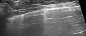
B mode: transthoracic view of lung (with linear probe)
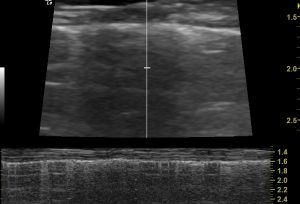
M mode: vertical lung pattern with fragmented pleural line is consistent with non-cardiogenic alveolar interstitial syndrome as described by Singh et al.

Detail of that M mode
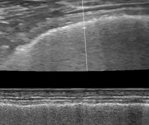
For comparison: transthoracic M mode of cardiogenic pulmonary oedema (CPO) from another canine patient

…and again, detail of the M mode
Early indications are that this looks really promising as a tool for us in veterinary medicine too.


