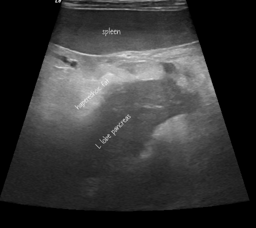
Sonography of canine acute pancreatitis: recent publications
Our understanding of the complexities of canine acute pancreatitis continues to develop through a revealing series of recent publications.
Secondary gastrointestinal changes in acute pancreatitis:
J Vet Intern Med 2022 May;36(3):947-956
Prevalence of ultrasonographic gastrointestinal wall changes in dogs with acute pancreatitis: A retrospective study (2012-2020)
Joshua J Hardwick, Elizabeth J Reeve, Melanie J Hezzell, Jenny A Reeve
https://www.ncbi.nlm.nih.gov/pmc/articles/PMC9151481/
‘Results: Sixty-six dogs were included. Forty-seven percent of dogs (95% confidence interval [CI], 35.0%-59.0%; n = 31) with AP had ultrasonographic gastrointestinal wall changes. Gastrointestinal wall changes were most common in the duodenum and identified in 71% (n = 22) of affected dogs. Of dogs with gastrointestinal wall changes, 74.2% (n = 23) had wall thickening, 61.3% (n = 19) had abnormal wall layering, and 35.5% (n = 11) had wall corrugation.’
J Vet Intern Med. 2019 Jan-Feb; 33(1): 79–88.
Computed tomographic angiography and ultrasonography in the diagnosis and evaluation of acute pancreatitis in dogs
John M. French, David C. Twedt, Sangeeta Rao and Angela J. Marolf
https://www.ncbi.nlm.nih.gov/pmc/articles/PMC6335445/
‘Evidence of gastric or intestinal wall thickening in the region of the inflamed pancreas was seen in 8 of the 26 dogs‘
Fig 4 of this paper shows dramatic images of a patient with histopathologically-proven necrotising pancreatitis and resulting duodenal infarction and perforation.
J Vet Intern Med 2019 May;33(3):1260-1265
Ultrasonographic features of presumed gastric wall edema in 14 dogs with pancreatitis
Masahiro Murakami 1, Hock Gan Heng 1, Chee Kin Lim 1, Nolie K Parnell 1, Nicholas J Rancilio 1, Tsang L Lin 2, Mario Sola 2
https://www.ncbi.nlm.nih.gov/pmc/articles/PMC6524385/
Speaks for itself: their findings are very similar to gastric wall oedema shown in our blog post here:
These studies out of Bristol, Colorado and Purdue reinforce the growing awareness of sonographic changes of the gastrointestinal tract which, we now know, are relatively common in pancreatitis cases. This is important because primary GI disease is an important alternative differential for abdominal inflammation and the resulting hyperechoic mesentery can occur in the same areas as those liable to be affected in pancreatitis.
Chronology of sonographic changes in acute pancreatitis:
Vet Sci. 2020 Dec; 7(4): 180.
Ultrasonographic Monitoring in 38 Dogs with Clinically Suspected Acute Pancreatitis
Federico Puccini Leoni, Tina Pelligra, Simonetta Citi,* Veronica Marchetti, Eleonora Gori, and Caterina Puccinelli
https://www.ncbi.nlm.nih.gov/pmc/articles/PMC7711604/
We really like this paper: it casts a whole new perspective on our approach to acute abdomen cases.
‘Dogs were submitted to two AUEs, the first on the first day of hospitalization, and the second between 40-52 h after the first one. Twelve dogs had both AUEs suggestive of AP. Fourteen dogs received an ultrasonographic diagnosis of AP exclusively on the second AUE. Twelve dogs remained negative on both the first and the second AUE. In 26 out of 38 patients the second AUE was suggestive of AP. If a patient is suspected of AP, it is advisable to carry out ultrasonographic monitoring at least within the first 52 h after admission, since ultrasonographic signs of AP may only become observable later after hospitalization.’
The important detail is that these dogs were scanned the first time within 6 hours of admission (time from onset of signs unknown) and again 40-52 hours later.
A picture is worth a thousand words; and the figures in this paper leave no doubt that we are missing a lot of pancreatitis cases if we judge them only on sonography at time of admission.
Similarly…..
J Am Vet Med Assoc 2021 Sep 15;259(6):631-636
Evaluation of diagnostic and prognostic usefulness of abdominal ultrasonography in dogs with clinical signs of acute pancreatitis
Eleonora Gori, Alessio Pierini, Ilaria Lippi, Simonetta Citi, Tommaso Mannucci, Veronica Marchetti
https://pubmed.ncbi.nlm.nih.gov/34448616/
‘24 of 37 (64.8%) dogs had AUS findings of AP at hospital admission, whereas [an additional] 10 had positive findings for AP on AUS within 2 days of hospitalization‘
It’s interesting that CT changes do not seem to evolve in the same way….
Vet Radiol Ultrasound 2020 Jan;61(1):33-39
CT angiographic changes in dogs with acute pancreatitis: A prospective longitudinal study
John M French 1, David C Twedt 2, Sangeeta Rao 2, Angela J Marolf
https://pubmed.ncbi.nlm.nih.gov/31596018/
CT vs US:
There’s still hope for sonographers….
J Vet Intern Med. 2019 Jan-Feb; 33(1): 79–88.
Computed tomographic angiography and ultrasonography in the diagnosis and evaluation of acute pancreatitis in dogs
John M. French, David C. Twedt, Sangeeta Rao and Angela J. Marolf
https://www.ncbi.nlm.nih.gov/pmc/articles/PMC6335445/
‘Computed tomographic angiography did not significantly identify changes indicative of pancreatitis, including pancreatic enlargement, mesenteric changes (P = .12), or changes in attenuation and echogenicity better than US (P = .14)‘
However, CT did reveal portal vein thrombosis in a whopping 9/26 cases….of which only one was picked up on US. My suspicion would be that sonographers did not specifically search for PV thrombosis…perhaps due to abdominal pain or because views of the PV were obscured by hyperechoic peripancreatic fat. This may be something that could be improved with a specific search in such cases. As the authors point out, detection of PV thrombosis has prognostic implications (longer hospitalisation).
cPLI accuracy:
This is a perennial debate but it’s worth noting that 8/26 were negative (cPLI<200; i.e. they would have had negative SNAP cPL test results) in the small case series of French et al. Obviously, plenty has been published on sensitivity and specificity of cPLI. It’s just worth reporting this particular finding because, in human medicine, Contrast-enhanced computed tomography (CECT) is the gold standard for ante-mortem diagnosis, I believe that French et al. represents the largest published case series on the subject in canine medicine to date.


