Sonographic features of cardiac hepatopathy in a dog with cor triatriatum dexter
I have to confess that this dog caught me out initially. A 12 m.o. Springer Spaniel; relatively well in herself but presenting with abdominal distention due to progressive ascites (of the modified transudate variety). No loud murmur.
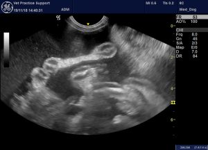
This is the liver:
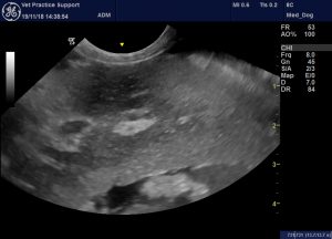
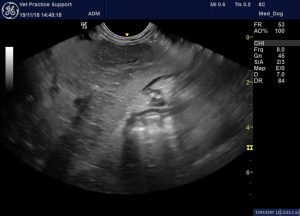
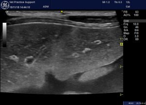
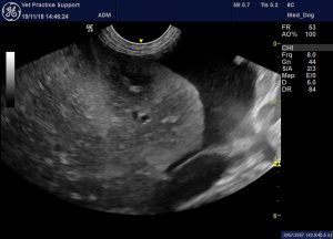
That’s not normal. The parenchyma is heterogeneous: specifically, the centres of all lobes are hypoechoic -contrasting with the periphery. A very unusual pattern!
Looks like a potential hepatopathy…Springers get chronic hepatitis….probably that…
But liver enzymes and bile acids normal….back to the drawing board
A closer look at the abdominal cava:
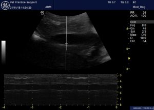
That doesn’t seem to be collapsing a lot with respiration.
Hepatic vein flow:
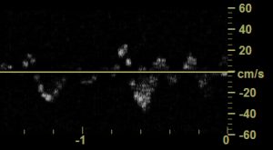
Interesting…normalish…but d waves are smaller than normal.
Anyway, certainly worth a proper look at the heart:
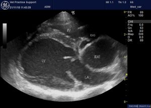
Cor triatriatum dexter.
An abnormal membrane is restricting flow from the coronary sinus and caudal vena cava into the main right atrium. The hole in the membrane is quite small:
That’s a transverse view:
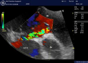
Anyway, so the cause of the ascites is post-hepatic venous obstruction between cava and RA. That’s a very similar phenomenon to Budd Chiari syndrome in man. And Budd Chiari syndrome in man is associated with a very interesting sonographic liver appearance.
Check out image A1 in:
https://www.ajronline.org/doi/10.2214/AJR.04.0918
American Journal of Roentgenology
July 2006, Volume 187, Number 1
Hepatobiliary Imaging
Pictorial Essay
Sonography of Budd-Chiari Syndrome
Xavier Bargalló1, Rosa Gilabert1, Carlos Nicolau1, Juan Carlos García-Pagán
Looks pretty much like our Springer’s liver.
Liver changes in human Budd Chiari involve central congestion (presumably the cause of central hypoechoic change) and progressive fibrosis (presumed cause of peripheral hyperechoic change) which can eventually progress to cirrhosis. Cardiac hepatopathy appears to be an interesting subject:
http://www.archivesofpathology.org/doi/pdf/10.5858/arpa.2015-0388-OA


hi, first of all thanks for this great page. We diagnosed CTD in one of my patients and we found evidence of ascites around the liver in that dog. What would you recommend as treatment options in such situations?
welcome, glad it was useful. Ideally surgery (balloon dilation) if you can find someone to do it. Otherwise palliative diuresis.