Sonographic features of septic arthritis in the dog
This is a nice article from a few years back:
BMJ. 2014 May 29;348:g3463. doi: 10.1136/bmj.g3463.
Will ultrasound scanners replace the stethoscope?
Wittenberg M.
https://www.bmj.com/content/348/bmj.g3463
The crux of it being that human medics are finding more and more advantages of using point-of-care ultrasound (POCUS) more as part of the physical examination, like a stethoscope, rather than as a ‘set piece’ diagnostic procedure. This is something which we would certainly advocate…but that’s another blog and current vet practice charging structures rather discourage this kind of thing.
Anyway, an interesting quote from that paper comes from a rheumaologist:

…which is interesting because there are quite a few settings in small animal practice where scanning joints is very useful. It’s much easier to be definitive about whether there’s a joint effusion when you can see it.
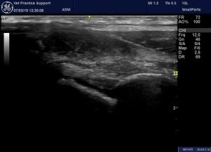
This is a recent septic shoulder in a Shar Pei:
The infraspinatus tendon crosses the middle of the image. On either side there is a joint effusion with markedly turbid, echogenic joint fluid. Surrounding soft tissues are hyperechoic.
Ultrasound-guided joint tap is the logical next step and this is much nicer than doing it ‘blind’.

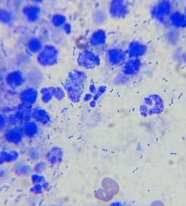
This is certainly septic: lots of cocci
Compared to the same area in a normal shoulder the contrast is striking:

Sterile inflammatory arthritis can obviously look rather similar: although the reaction in surrounding tissues tends to be less dramatic.


