Sonographic features of canine shoulder osteochondrosis (OCD)
I have blogged about this before:
However, at that stage I hadn’t found this:
Vet Radiol Ultrasound. 2015 Jan-Feb;56(1):3-11. doi: 10.1111/vru.12179. Epub 2014 May 21.
Diagnostic sensitivity of radiography, ultrasonography, and magnetic resonance imaging for detecting shoulder osteochondrosis/osteochondritis dissecans in dogs.
Wall CR1, Cook CR, Cook JL.
https://www.ncbi.nlm.nih.gov/pubmed/24844132
Which, happily, demonstrated that ultrasonography was broadly comparable to radiography in the detection of shoulder OCD lesions.
‘Diagnostic sensitivity, specificity, and accuracy (correct classification rate) values for detecting presence or absence of shoulder osteochondrosis/osteochondritis dissecans were as follows: radiography (88.5%, 90%, 88.9%), ultrasonography (92%, 60%, 82.6%), and MRI (96%, 88.9%, 94.4%)‘
Sonography is obviously highly dependent on machine/probe specifications and technique. In an exciting development, we are proud to include the amazing images of Bob Hylands from Ontario acquired using a Logiq S8 linear matrix probe: they are the best I’ve seen.
http://www.westbridgevet.com/about/our-veterinarians
I
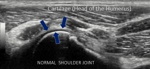
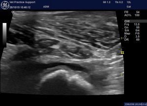
Oblique view of the caudolateral aspect of a normal canine humeral head. Note the physis: which could be mistaken for a pathological defect (Bob Hylands).
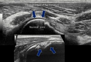
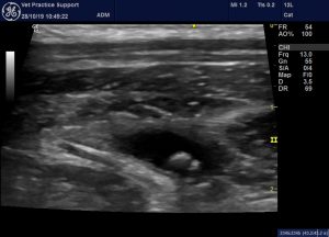
…and in comparison with a shoulder bearing an OCD lesion (Bob Hylands)
This is one of mine (Logiq R7 linear probe):
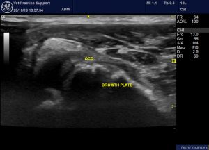
Same view showing relationship between OCD lesions and growth plate.
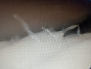
This is the appearance of that OCD lesion at arthroscopy.
Images courtesy of Bob Pettit at Kingsway Vets, Skipton
The growth plate in more detail (there is a joint effusion):
This dog does have OCD, hence the joint effusion, but the feature in this image of the humeral head is the normal growth plate.
Joint ‘mice’ in the caudal pouch of the shoulder joint in the same dog.
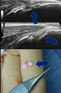
The flap retrieved at surgery (Bob Hylands).
And finally,
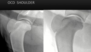
Radiographic appearance: shows the relationship between the physis and OCD lesion (Bob Hylands)


