Sonographic features of bacterial discospondylitis in a dog
I’m not suggesting that ultrasound should necessarily be your imaging modality of choice when suspecting discospondylitis; but, as always, these cases don’t always arrive with a label. The point is that if one is triage-ing a febrile or paraplegic patient with ultrasound then it’s good to extract maximum information whilst one has the chance. It’s easy enough to see the ventral aspect of at least some of the vertebrae and intervertebral discs when performing abdominal ultrasound and there are often changes in the retroperitoneal soft tissues when there’s pathology of the lumbar spine.
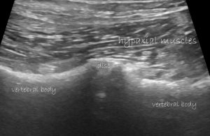
oblique, parasagittal plane view of the hypaxial muscles and ventral aspect of lumbar vertebrae with a single disc visible in this field of view
This image is a healthy area of lumbar spine from a previously healthy Springer Spaniel (obviously it’s a Springer!) presenting with acute onset fever, symmetrical paraplegia, malaise and anorexia.
In the sublumbar, retroperitoneal soft tissue there are small pockets of free fluid.
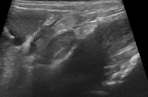
Looking a bit further in this area, the small effusion and associated hyperechoic soft tissue are focused on the ventral aspect of L3, specifically the cranial end-plate.
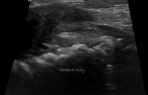
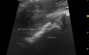
Parasagittal plane view of the ventral aspect of L3 and adjacent disc. The substance of the intervertebral disc is markedly heteroechoic
There’s a lot of proliferative periosteal new bone along the underside of the vertebral body.
Furthermore, there were no changes elsewhere in the heart, thorax or abdomen to explain his current presenting signs.
On radiography, the same changes are visible:
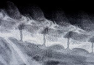
On the basis of a presumptive diagnosis of discospondylitis (+/- foreign body) treatment was started with coamoxiclav. Prompt improvement in all signs was observed with return to normal within a week.


