Sonographic diagnosis and surgical management of perforating, benign duodenal ulcer in a dog
Gastric and duodenal ulceration is probably relatively common in dogs: usually secondary to NSAID and/or glucocorticoid treatment, liver disease, hypoadrenocorticism, mast cell tumours or gastrinoma. However, perforation and subsequent septic peritonitis is less frequent. In fact there are few published cases.
Hinton L. E., McLoughlin M. A., Johnson S. E. & Weisbrode S. E. ( 2002)
Spontaneous gastroduodenal perforation in 16 dogs and seven cats (1982-1999) .
Journal of the American Animal Hospital Association 38, 176- 187
http://www.jaaha.org/doi/pdf/10.5326/0380176
Vet Rec. 2009 Oct 10;165(15):436-41.
Spontaneous gastroduodenal perforations in dogs: a retrospective study of 15 cases.
Cariou M1, Lipscomb VJ, Brockman DJ, Gregory SP, Baines SJ.
https://veterinaryrecord.bmj.com/content/165/15/436
In the latter series ‘…there was not one single investigative test that was diagnostic for gastroduodenal perforation before surgery in this study. Survey and contrast radiography of the abdomen, ultrasonography and, most importantly, abdominocentesis and cytological evaluation were all helpful for increasing clinical suspicion of gastroduodenal perforation.‘
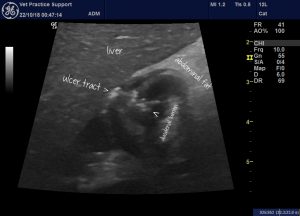
In a cursory search of the internet, texts and scientific journals I was also unable to find an image of a perforating duodenal ulcer in a dog. My suspicion is that’s because, predominantly, these are large breed dogs with musculoskeletal issues necessitating NSAID use. Large breed dogs are much more difficult to scan in detail: and in particular, the pylorus and cranial duodenal flexure are relatively inaccessible beneath the rib cage.
However, it is possible to visualise duodenal ulcers sonographically and very helpful to do so since this facilitates a rapid decision for surgery without further interventions. Frequently, the best views are obtained with transverse or oblique planes of imaging through the intercostal spaces. The optimal contact point is often very circumscribed.
The present patient is a 1 y.o. MN Labrador with a history of NSAID and glucocorticoid use (not concurrently) presenting with acute collapse, tachypnoea, fever and abdominal pain. This is a transverse view of the proximal duodenum:
…and a frozen image of the same area:

As always with these things, the key to the crux of the problem is localising the focus of hyperechoic change in the abdominal fat in a rapid intial sweeep of the abdomen. Although the right cranial abdomen was clearly most severely affected it was also apparent that the specific focus was the pyloric area rather than pancreas or gallbladder.
At exploratory laparotomy a small perforating ulcer of the anti-mesenteric wall of the duodenum was confirmed.
The literature also offers little objective evidence to support one surgical option over any other in such circumstances. Cariou et al. opted to debride and suture most cases with Billroth I procedures performed in more extensive cases. In human medicine it appears that simple closure is also preferred in most cases:
‘Most authors recommend simple oversewing of the ulcer in addition to treating the underlying Helicobacter pylori infection or cessation of nonsteroidal anti-inflammatory drugs (NSAIDs)‘
https://emedicine.medscape.com/article/1950689-overview#a4
Happy to report that, after oversewing, this particular patient went on to recover uneventfully.


