Sonographic assessment of oesophageal function in dogs
I am starting to think that examination of the neck might be a useful part of the routine sonographic examination for cats and dogs: especially in complex cases. In addition to those with sepsis, foreign bodies, thyroid disease, lymphadenopathy or salivary gland changes, patients with impaired oesophageal function are sometimes picked up. Some animals with oesophageal dysfunction can be quite cryptic; they present with cough, fever, inappetence, dysphagia, weight loss and other not-very-specific signs. Or the oesophageal issue may be part of a wider spectrum of myopathy and can get missed.
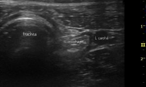
This is a longitudinal plane view of the cervical oesophagus in an unsedated dog in standing position with polymyositis and enteric ganglionitis who presented intially with inability to close his jaw and then a few months later with a dysautonomia-like syndrome of global gut hypomotility.
There is an abnormal, persistent accumulation of liquid saliva/ingesta in the oesophagus above the thoracic inlet. The oesophageal lumen should normally be empty.
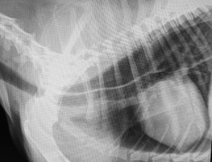
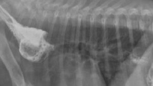
On radiography there is a convincing thoracic megaoesophagus.
And, intermittently, dilation of the small intestine with gas:
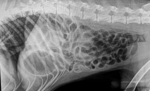
Histopathology demonstrated a ganglionitis in the abdominal GI tract wall.
Another case: this is a Viszla dog with polymyositis:
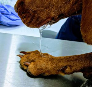
Sialorrhoea is a common sign of polymyositis in Vizslas
This is an excellent account of the disease in this breed specifically:
Clinical features of idiopathic inflammatory polymyopathy in the Hungarian Vizsla
Anna Tauro, Diane Addicott, Rob D Foale, Chloe Bowman, Caroline Hahn, Sam Long, Jonathan Massey, Allison C Haley, Susan P Knowler, Michael J Day, Lorna J Kennedy & Clare Rusbridge
BMC Veterinary Research volume 11, Article number: 97 (2015)
https://bmcvetres.biomedcentral.com/articles/10.1186/s12917-015-0408-7
In cases with unexplained pneumonia, dysphagia, sialorrhoea and in cases where it’s difficult to say whether the ‘bringing up food’ is vomiting or regurgitation then ultrasonography of the cervical oesophagus is a quick and convenient screening tool to assess the likelihood of oesophageal dysfunction -especially in circumstances where it seems a bit of an over-reaction to go straight for barium.
This is a transverse view of the neck (from the ventral aspect) in a dyspnoeic 10 w.o. pup:
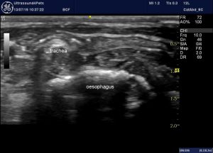
Lung ultrasound revealed typical ‘shred sign’ of bronchopneumonia. The cervical oesophagus, seen here in transverse view is hugely dilated with gas/ingesta.
Transverse view of a healthy dog’s oesophagus for comparison:
The oesophageal lumen should normally be empty or nearly so
Abrupt constriction of the oesophageal lumen at the heart base consistent with vascular ring anomaly was confirmed on contrast radiography:
In idiopathic megaoesophagus the thoracic oesophagus is often dilated to the extent that it contacts the chest wall in the cranial thorax. This is a transthoracic view at the second intercostal space:
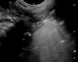
Longitudinal plane view from the second intercostal space: the oesophagus is hugely dilated with saliva/ingesta.


