Sonographic appearance of pancreaticoduodenal lymph nodes in dogs and cats
Sonographic heterogeneity of abdominal lymph nodes has been reported as a feature associated with malignancy in dogs:
Vet Radiol Ultrasound. 2007 Nov-Dec;48(6):565-9.
Association between malignancy and sonographic heterogeneity in canine and feline abdominal lymph nodes.
Kinns J1, Mai W.
https://www.ncbi.nlm.nih.gov/pubmed/18018731
In that series, 91% of dogs with heterogeneous abdominal nodes proved to have malignancies.
However, the principal pancreaticoduodenal node is an exception to this rule: being consistently heterogeneous in both cats and dogs under normal circumstances.
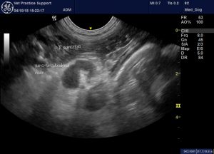
Longitudinal plane view from the right flank showing the pancreaticoduodenal lymph node in a dog with acute pancreatitis (hence the hyperechoic perinodal fat)
There appears to be a ‘finger’ of abdominal fat indented into the hilus of the node which creates this appearance.
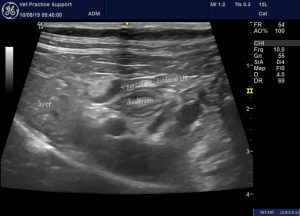
Similar view from a cat with an acute enteritis. The pancreaticoduodenal node may be somewhat enlarged. It has the same apparent fat ‘indent’ as seen in dogs.
That’s the kind of ultrasound geekery we love 🙂


