Septic thrombus of the renal vein and vena cava in a Pug dog with pyelonephritis
This Pug presented semi-recumbent and febrile with severe abdominal pain.
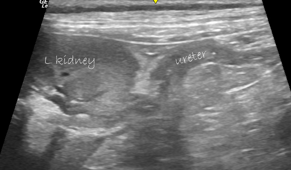
On abdominal ultrasound, the dominant pattern of change involved hyperechoic peri-renal retroperitoneal fat on the left side and small pockets of peri-renal and peri-ureteral effusion. The left kidney was diffusely hyperechoic with reduced cortico-medullary distinction but of unremarkable overall size and shape. Neither renal pelvis nor proximal ureter were dilated but the wall of both and the surrounding fat exhibited increased echogenicity.
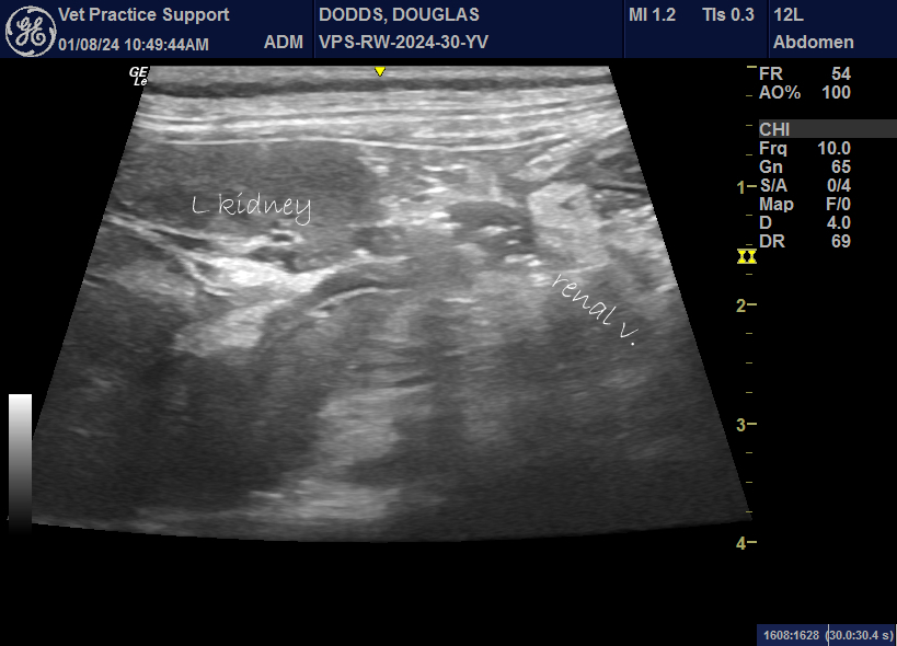
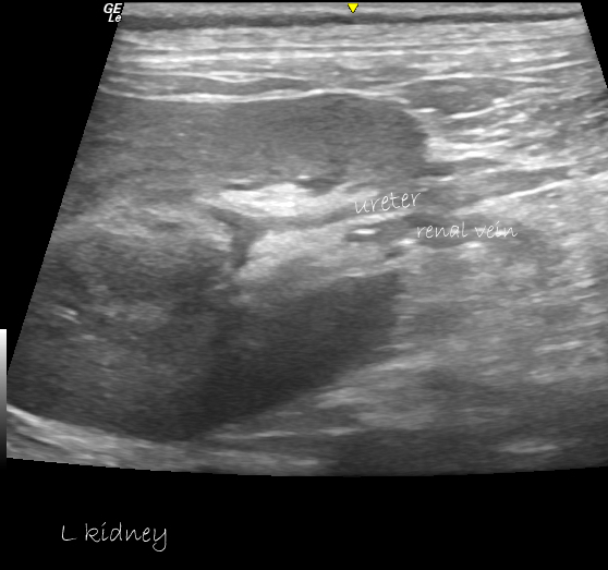
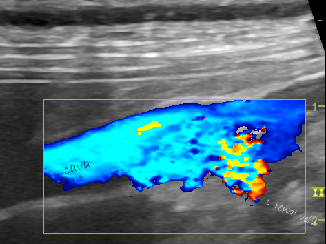
No uroliths were apparent. However, the lumen of the renal vein was, strikingly, studded with intensely hyperechoic non-shadowing foci (consistent with gas densities).
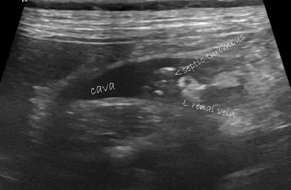
At the confluence with the abdominal vena cava, a presumed thrombus, also speckled with gas densities, dangled into the caval lumen.
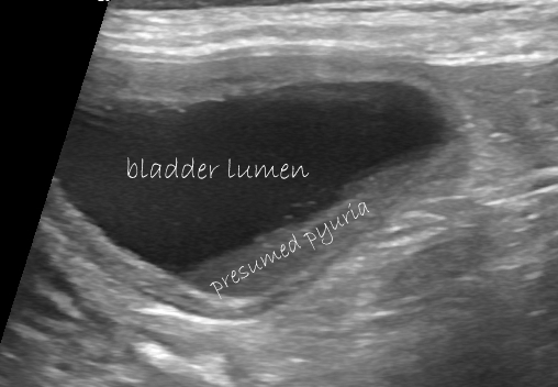
The bladder contained a thick drift of non-mineral sediment in the dependent part. urine sediment microscopy revealed abundant bacterial rods, red cells and neutrophils.
Lung ultrasound findings were unremarkable.
Sadly, despite medical management the patient deteriorated rapidly and his owners opted for euthanasia.
In a brief survey of the published literature, I can’t immediately find any previous cases of septic venous thrombus in dogs.
Even in human medicine it appears to be very rare:


