‘Renal FIP’: sonographic features
There are still relatively few peer-reviewed publications documenting sonographic changes associated with FIP in domestic cats.
J Am Anim Hosp Assoc. May-Jun 2010;46(3):152-60.
Abdominal ultrasonographic findings associated with feline infectious peritonitis: a retrospective review of 16 cases
Kristin M Lewis 1, Robert T O’Brien
Lewis and O’Brien remains the most comprehensive account. The only caveat is that it goes without saying that 16 cats can’t really do justice to the vast range of possible changes in such a protean disease. Obviously there are numerous mentions of imaging findings elsewhere in the literature.
These include echocardiographic changes in some cases:
https://www.ncbi.nlm.nih.gov/pmc/articles/PMC6787879/
https://www.jstage.jst.go.jp/article/dobutsurinshoigaku/25/4/25_148/_article/-char/en
https://www.koreascience.or.kr/article/JAKO201708260282817.pdf
The spectrum of sonographic findings which might be expected can be inferred from accounts of post-mortem and histopathological findings….which involve almost every organ!
The Advisory Board on Cat Diseases (ABCD) website seems to be pretty much the state of the art:
http://www.abcdcatsvets.org/feline-infectious-peritonitis/
The latest update March 2021 by Severine Tasker et al. includes a reference to the occurrence of renomegaly in some FIP cases and an ultrasound image.
Recently we have seen two cases in which the dominant pattern on sonography was of severe bilateral nephropathy with renomegaly and retroperitoneal effusion and/or renal subcapsular free fluid. Neither cat was azotaemic.
| Parameter | Case A | Case B |
| Age | 9 months | 13 months |
| Signs | fever, hyporexia | fever, uveitis, icterus, |
| albumin:globulin | 0.28 | 0.27 |
| FCoV titre | >1280 | >1280 |
| alpha-1 AGP | >5000 | |
| Effusion FCoV PCR | moderate positive | moderate positive |
| Renal cytology | No evidence lymphoma |
These are the kidneys in case B:
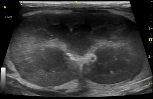
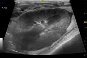
Dominant changes are renomegaly, retroperitoneal effusion, small amount of subcapsular fluid and diffuse cortical hyperechoic/heterochoic change.
This video clip is also case B and gives a better idea of the volume of retroperitoneal effusion:
There were no abdominal or thoracic effusions. Liver was diffusely hypoechoic compatible with acute hepatitis. Abdominal nodes were not markedly enlarged.
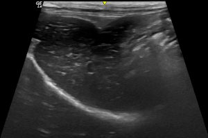
Case B: longitudinal plane view of liver exhibiting diffuse hypoechoic change. There is no abdominal effusion
Case A showed more severe renal changes:
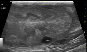
Case A: R kidney
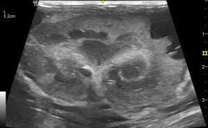
Case A; L kidney
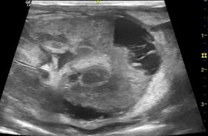
Case A: L kidney focussing on one subcapsular cystic area
In this case there was more destruction and distortion of normal kidney anatomy: largely due to multiple subcapsular cystic areas. Retroperitoneal effusion, diffuse heterogenous hyperechoic change in the cortices were, again, striking features.
Again there were no abdominal or thoracic effusions. A scatter of consolidated lung lesions were present consistent with pneumonitis.
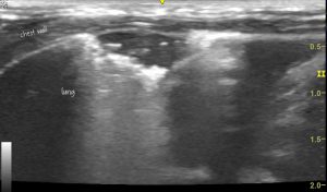
Case A: lung consolidation. There were numerous such areas with characteristics of ‘shred sign’ or ‘fractal sign’ suggesting pneumonia/pneumonitis. This is quite a common finding in FIP.
Case A video clip R kidney:
Both cats were euthanased.
These cats, along with others with echocardiographic changes, have forced us to reassess our diagnostic criteria for potential FIP in our patients presented for sonography. It’s increasingly apparent that FIP can be very variable. Whilst ultrasound is a very valuable tool in all of these cats and can help build a case for a diagnosis of FIP, we are more and more aware that many cases don’t fit any ‘classic’ pattern -especially in earlier stages.
As the ABCD website says ‘The distinction between so-called ‘effusive’ and ‘non-effusive’ forms of FIP is important for diagnostic purposes; however, there is considerable overlap between the two forms, and indeed FIP cases with effusions also have pyogranulomatous lesions visible at post-mortem examination and, similarly, many cats with a non-effusive form will eventually develop effusions‘.
To add to this we now have cats recovering or recovered after remdesivir or GS-441524 treatment……


