Re-formation of the gallbladder after cholecystectomy in a dog
An 11 y.o. FN CKCS was examined for investigation of multiple problems including degenerative mitral valve disease, splenic mass, pancreatitis and urinary tract infection. She had already undergone cholecystectomy via laparotomy several years earlier for management of gallbladder mucocoele.
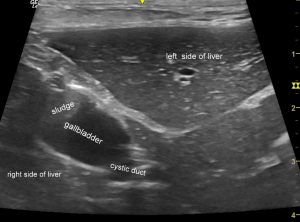
We were surprised to find that there was a relatively normal-looking gallbladder in a conventional position right of midline. The lumen containing anechoic bile and a small amount of mobile sludge.

Transverse plane view of the liver from close to the xiphisternum. A slightly small but otherwise fairly unremarkable gallbladder is present and continues, as expected, into the cystic duct.
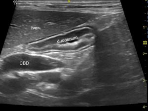
The remainder of the extra-hepatic biliary tract was moderately dilated down to the level of the papilla with no obvious obstruction.

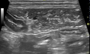
Although the swollen, marbled appearance of the pancreas suggests the possibility of some (likely chronic) pancreatitis which might cause occlusion or fibrosis of the distal bile duct.

There is an interesting report in the human medical literature:
https://academic.oup.com/jscr/article/2015/2/rju154/2412602
Reformed gallbladder after laparoscopic subtotal cholecystectomy: correlation of surgical findings with ultrasound and CT imaging
Suzanne J. Di Sano Nicholas B. Bull
Journal of Surgical Case Reports, Volume 2015, Issue 2, 1 February 2015,
…which essentially reports the same thing.
Post-cholecystectomy syndrome is a well-documented phenomenon in people. It is hypothesized that once the reservoir of the gallbladder is removed, any spasm or fibrosis of the sphincter of Oddi readily results in increased biliary pressure.
https://www.ncbi.nlm.nih.gov/pubmed/6479537
Gastroenterology. 1984 Nov;87(5):1154-9.
Change in bile duct pressure responses after cholecystectomy: loss of gallbladder as a pressure reservoir.
Tanaka M, Ikeda S, Nakayama F.
It seems possible that this might be responsible for a progressive dilation of the remnant cystic duct to form a new gallbladder or pseudo-gallbladder.


