Pulmonary thromboembolism in a dog
I’m interested in lung ultrasound. I feel it has a lot to offer, it’s quick and easy. For a while I’ve been waiting for a convincing case of pulmonary thromboembolism (PTE) to come along. It seems very likely that such cases exist amongst our workload but, since we don’t do a lot of CT angiography, I presume we are missing them. The veterinary literature has very little on the sonographic appearance of PTE. In human medicine PTE is usually described as causing discrete, often wedge-shaped, subpleural lesions.
Today’s patient then is a middle aged Greyhound with a history of chronic protein-losing nephropathy, hypoalbuminaemia and systemic hypertension. She was relatively stable until an acute deterioration with malaise, coughing and inappetence 48 hours prior to sonographic examination.
On physical examination she had dull percussion in the cranial and ventral left thorax and a new grade III systolic right-sided murmur.
The dull area looks like this on transthoracic ultrasonography:
There is patchy consolidation of the subpleural lung and increased B lines (ultrasound lung rockets, comet-tail artefacts) But the whole right hemithorax and the remainder of the left side look pretty unremarkable:
Here the A lines (parallel to the pleura) are visible as normal.
Differentials for the appearance of the abnormal lung include PTE, contusions and pneumonia. Cardiogenic pulmonary oedema can be asymmetrical but I don’t believe it is ever likely to be this localised; nor is this degree of consolidation (hypoechoic areas).
And now echocardiography. This is a left apical four-chamber view:
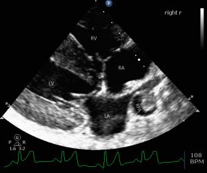
The right heart is clearly dilated but right ventricular hypertrophy is absent.
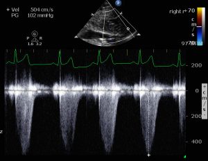
Tricuspid regurgitation is present and maximum velocity is a whopping 5m/s:

Equating to pressure gradient of 125mmHg. Obviously, even if RA pressure were zero this would indicate a profoundly supranormal RV systolic pressure -and since, in systole, the pulmonic valve is open, pulmonary arterial pressure must be the same.
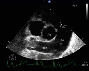
In a right long axis four-chamber view the dilation of the right heart is also apparent. Moreover, there is an abnormal structure in the right main pulmonary artery:

This is confirmed in a transverse view:

Given that this is acute pulmonary arterial hypertension (PAH) it’s difficult to imagine this being anything other than a thrombus. The only alternative being, theoretically, a neoplasm.
Protein-losing nephropathy is a known predisposing factor for thromboembolic disease generally.


