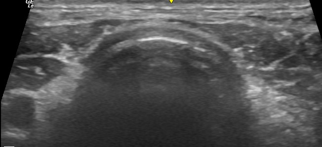
Practical small animal ultrasound bits and pieces: part II…. tracheal exudate sign
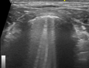
Same view in a coughing dog. There is frothy exudate on the internal aspect of the tracheal wall: bubbles trapped within mucus/exudate cause reverberation artifact ‘comet tails’.
And the same in video:
This is a useful finding: it implies airway exudate. If you see this in a coughing patient and transthoracic lung ultrasound is unremarkable, there is likely significant airway disease. Differential diagnoses associated with this might include, for example, eosinophilic bronchopneumopathy, infectious tracheo-bronchitis, bleeding due to coagulopathy, bronchial foreign body.
Also another good reason to scan the neck in every patient! Always useful to have a handle on the appearance of the trachea, thyroids, parathyroids, cervical nodes and oesophagus.


