Practical small animal ultrasound bits and pieces: part I…linear foreign bodies
Linear FBs are sometimes difficult to visualise convincingly: even when everything is plicated to hell.
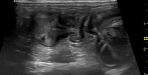
Plicated small intestine in the abdomen of a cat with a history of several days’ vomiting and deteriorating malaise. OK, we might opt to open that one up on the basis of plication alone: but it’s a lot more satisfactory to be definitive about the cause.
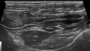
The same cat; another area of jejunum. Is that a linear thing in the lumen there? Hairs are commonly seen in the gut lumen though: and they look a lot longer on ultrasound than you expect them to be!
OK, now see what happens if you fan the probe over the suspect loop of gut…
…like magic! There it is.
One more still from the same cat. We know the linear FB is there:
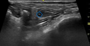
In still images, the linear FB is often just seen as an isolated hyperechoic focus where the plane of scanning cuts through it.


