Potential haemocholecyst in a young dog with acute leptospirosis
The patient in this case is a 6 m.o. Labrador pup who presented with acute severe fever, malaise, vomiting, melaena and jaundice.
Blood revealed raised bilirubin, alkP, ALT, urea, creatinine and phosphate, isosthenuria, glycosuria and mild anaemia.
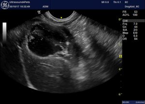
An initial scan by the primary care vets revealed no specific abnormalities other than a very abnormal-looking gallbladder. Two days later we had the opportunity to have a look ourselves:

An oblique view of the gallbladder from the right ventral abdominal wall
The gallbladder is largely filled with hypoechoic ‘material’. But it’s not bile. The hypoechoic stuff is immobile and contains hyperechoic flecks and strands.
Under other circumstances the initial reflex would be to assume that the hypoechoic material might be mucus. At least superficially it does look a bit like a mucocoele…..but in a 6 month old dog! And if this were a rupturing mucocoele then why is there no hyperechoic reaction in the fat around the porta hepatis?
Now take a look at the video:
On closer inspection the hypoechoic material is an irregular, flexible lump of stuff ‘floating’ within the lumen and surrounded by mobile bile. The gallbladder wall is actually pretty unremarkable: thin and even. There is no effusion or reaction around the outside of the gallbladder.
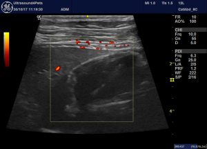
The ‘mass’ is not vascularised:

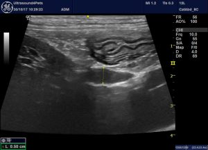
The common bile duct is mildly dilated in its proximal part and appears to be full of hypoechoic solid matter:

longitudinal view from the right flank showing pyloric antrum and common bile duct. Note that the fat around the duct is not hyperechoic (no evidence of pancreatitis or bile peritonitis).
So, what the blazes is going on here???? What is the hypoechoic stuff and how does it relate to the dog’s signs and blood biochemistry findings?
My theory is that it’s a blood clot. This would be a phenomenon known as a haemocholecyst. There appear to be just a couple of reports in the veterinary literature. Both were associated with neoplasia of the gallbladder wall and were presumably somewhat more chronic than the present case.
Acute hemobilia and hemocholecyst in 2 dogs with gallbladder carcinoid.
An ultrasonographic image comparable to our case can be seen in this human case:
https://synapse.koreamed.org/Synapse/Data/PDFData/0028KJG/kjg-54-63.pdf
The primary cause in that case being ‘ 초음파검사에서 담낭의 확장과 담낭벽 비후 소견을 보였고 확장된 담낭 내에 불규칙한 모양의 고음영 종괴가 관찰되었으며 내부는 다양한 음영의 층’
(apologies to Korean readers!)
Our dog turned out to have a strong positive IFA test result for Leptospira spp: an outcome consistent with a presentation involving apparent acute kidney injury, hepatocellular damage and potentially with coagulopathy (although no coagulation parameters were measured during the acute phase). He made an uneventful recovery within a week after supportive treatment and antibiotics.


