Portosystemic shunts involving the left phrenic vein in cats
Our understanding of shunt anatomy in cats and dogs has moved forward recently with advances in imaging technology:
http://www.ncbi.nlm.nih.gov/pubmed/26563977
http://www.ncbi.nlm.nih.gov/pubmed/23888909
In cats, shunts through the right gastric vein appear not to have reported; whereas in dogs they are quite common. Broadly-speaking, feline congenital extrahepatic shunts may originate at 1) splenic vein 2) mesenteric vein or 3) colic vein sites.
Most anomalous vessels arising from the base of the splenic (or gastro-splenic) vein have been shown to flow in a hepatopetal direction through the left gastric vein -which is normally a tributary to the splenic vein just before it enters the main portal vein. This reversed flow is carried to join the systemic circulation at either 1) the cava 2) left phrenic vein or 3) azygous vein. Azygous vein shunts appear to be exceptionally rare in cats.
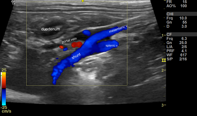
These are some images from a cat with a left gastric -left phrenic PSS. In normal cats the splenic and mesenteric veins join to form the portal vein and the left gastric is a smaller tributary to the splenic just before the junction.

longitudinal plane view from the right flank just caudal to the last rib: cranial is to the left of the image
The portal vein itself is abnormally narrow as most blood is diverted through the shunt.
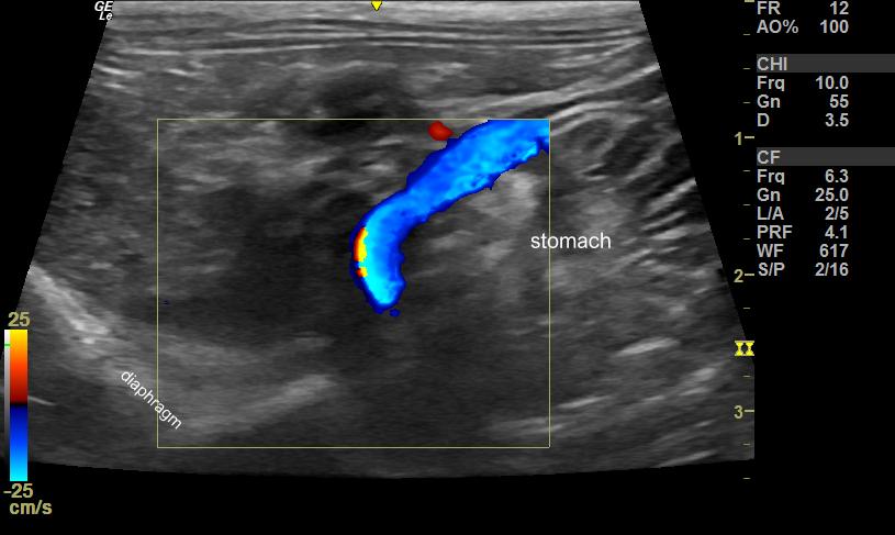
The shunt then dives cranio-dorsally across the lesser curvature of the stomach:

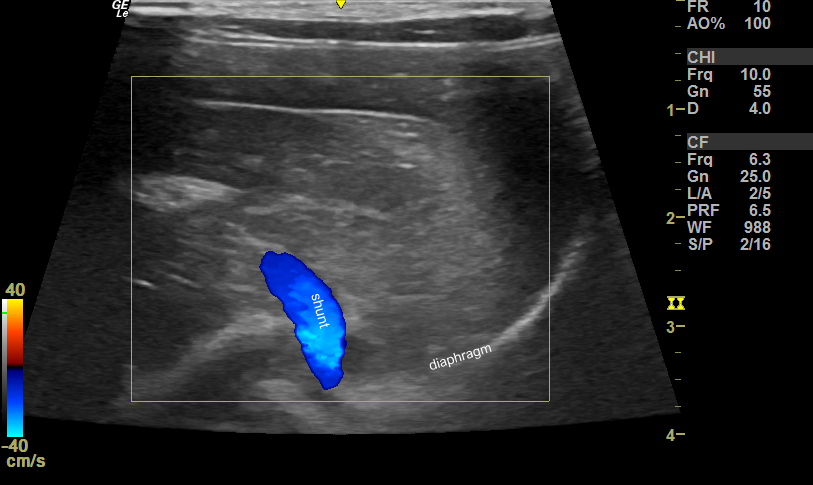
In transverse plane the shunt can be seen sliding to the left of midline:

transverse plane view: left of patient to right of image
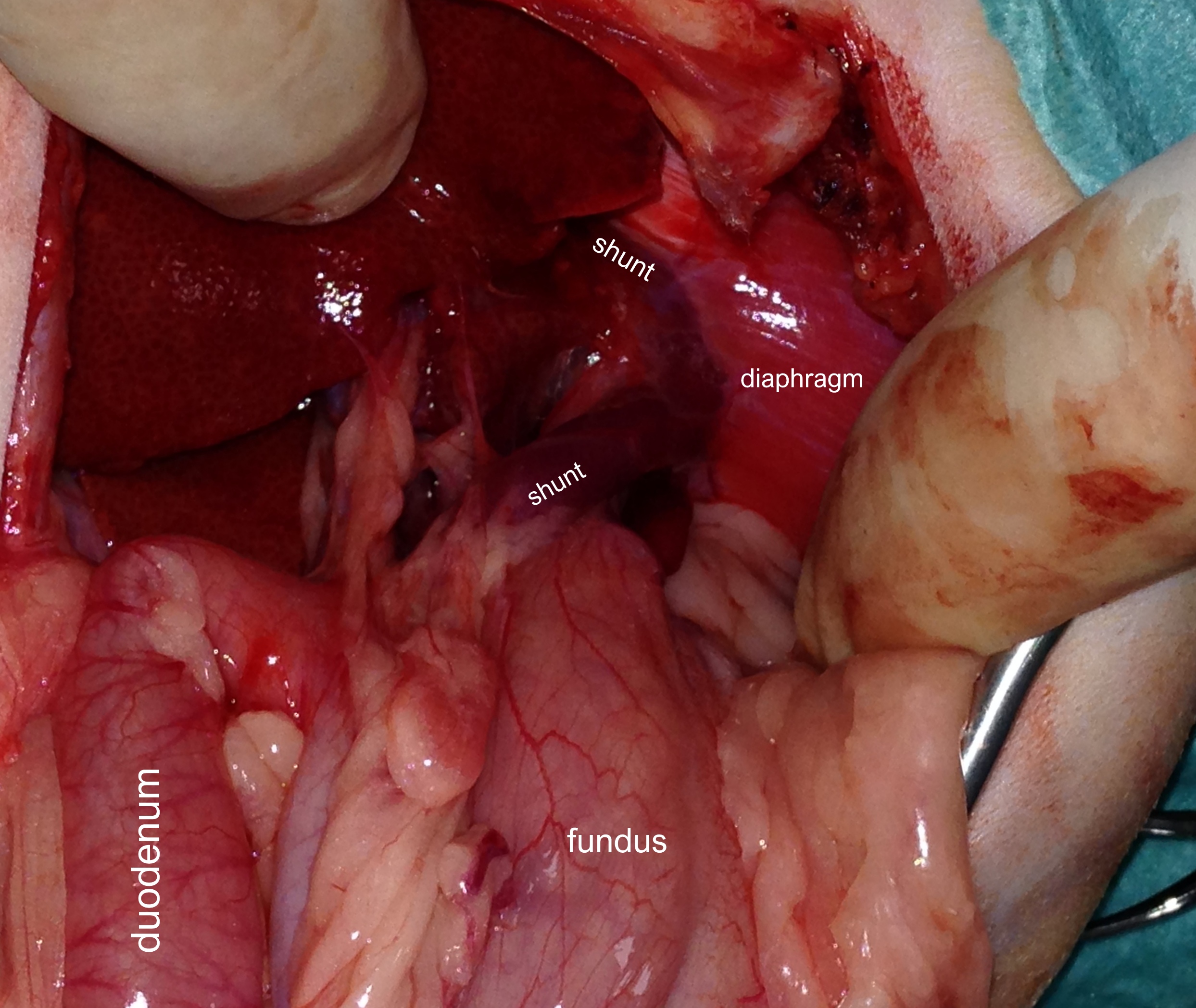
At surgery, it was confirmed that the anomalous vessel joins the left phrenic vein at the diaphragm. Flow then deviates 90 degrees into the phrenic vein and thence joins the cava

This arrangement of vessels facilitates the surgical approach as visualisation is reasonably straightforward.


