Portal vein anatomy in cats and dogs
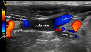
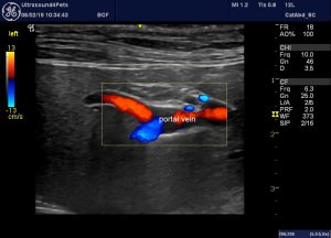
Just a little note with a couple of things that I’ve not seen mentioned elsewhere. A standard view of the portal vein as seen in longitudinal plane from the right side has an ‘envelope’ configuration:

In all of these images cranial is to the left of picture:

Canine portal vein in longitudinal plane as seen from the right, with colour Doppler
In this video you can also see the confluence of cranial and caudal mesenteric veins to the right of picture -before the splenic vein joins from the left (bottom of image).
It’s important to be able to identify the tributaries and branches with confidence so that when faced with a potentially abnormal arrangement you know what’s what.
For example, as you follow the portal vein forward into the liver it splits into left and right main branches. It’s not so difficult to mistake the right branch (again, seen from the right side) for the gastroduodenal vein. And with colour Doppler the flow would appear hepatofugal if one were to make that mistake.

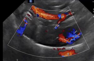
Superficially that could look like an extrahepatic shunt through the right gastric vein:

Right-sided longitudinal plane view of the cranial part of the portal vein in a dog with a congenital extrahepatic portosystemic shunt through the right gastric vein (the R gastric vein joins the pancreatico-duodenal vein just before the portal vein to form the gastroduodenal vein)
The difference lies in where the fork lies in relation to the porta hepatis and duodenum.
It’s always possible to identify the mesenteric vein because, following it caudally, the cranial mesenteric will run between the jejunal lymph nodes.
Similarly, the gastroduodenal vein can be followed back from the portal vein into the body of the pancreas (this can be a good way to identify the pancreas with certainty).
However, cats and dogs differ slightly in their gastroduodenal vein anatomy. In cats the vein usually appears to enter the portal vein more caudally in relation to the cranial duodenal flexure:
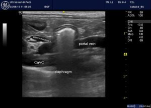
Whereas, in the dog, it enters cranial to the duodenum:

The exact appearance does depend on the angle of view. You’ll notice that in some of the videos the canine gastroduodenal vein does look to run caudal to the duodenum.


