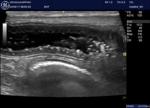
Pneumatosis in canine chronic enteropathy
Pneumatosis describes the presence of gas within gut wall of presumed bacterial origin.
In cats and dogs this is usually associated with acute gut wall necrosis such as haemorrhagic diarrhoea syndrome or gastric dilation-volvulus.
However, it can also be seen in chronic enteropathies. This is an elderly Westie with chronic weight loss and gastrointestinal signs. At the time of examination he was outwardly well.

long axis view of a loop of jejunum mid-abdomen
There are intensely echogenic foci within the mucosa. The main differentials for this appearance are dilated lacteals (as in lymphangiectasia) and pneumatosis. These differ subtly in appearance. Lacteals are often linear (perpendicular to the long axis of the gut) and the foci tend to be pleiomorphic and of variable size. The gas bubbles of pneumatosis, on the other hand, are usually more echogenic with dirty shadowing, non-linear and tend to be all of similar size.
In human medicine a wide variety of conditions are associated with pneumatosis:
http://www.ajronline.org/doi/full/10.2214/AJR.06.1309
These can be divided into groups 1) bowel necrosis 2) increased permeability 3) mucosal disruption 4) pulmonary disease.
The list includes inflammatory bowel disease and gastrointestinal lymphoma which are possibilities in this case.


I’m not sure if I respectfully agree with the Dx of pneumatosis especially when compared to the CT case examples. I suspect that you may just as likely be capturing the presence of gas bubbles trapped within the villi like projections of the mucosal surface of the jejunum. If you look closely they fit with the usual depth of that normal anatomy. They do not reach the sub mucosa layering. I wish that I could attach histology images to make my point. Perhaps I can send them out by email, sincerely, Bob.
Love your work ups.
Thanks Bob, you know what?….I think you may well be right. I was thinking about it a bit even as I was writing it. And since I’ve been thinking about it I’ve been looking more critically at hyperechoic foci in gut walls in subsequent cases. If you’ve got histo images by all means email us ultrasound4pets@gmail.com and I’ll put them up with a bit more discussion. All the best, Roger