Percutaneous ultrasound-guided biopsy of bone tumours in dogs
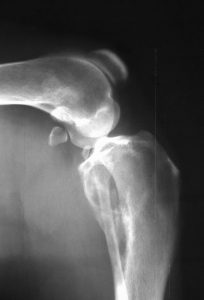
This is a radiograph of the stifle of a dog with a two-week history of progressive, severe lameness:

What do you think?
In fact, whatever you think, I’m betting you’re not 100% certain. Now have a look at the ultrasonographic images:
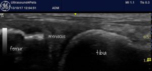
This is the medial aspect of the stifle joint with the tibia to the right:

And that’s how a long bone should look: a smooth cortical surface and virtually no sound penetration beyond that interface.
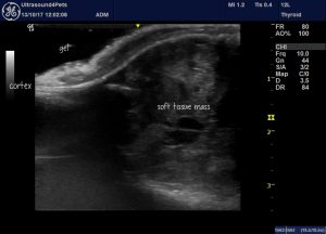
On the lateral aspect of the proximal tibia it looks like this:

Now I am 99% certain that this is a neoplastic lesion and the examination took about 20 seconds. There is osteolysis of the overlying cortex to the extent that the marrow cavity below is clearly visible. The cavity is filled with abnormal soft tissue. The adjacent cortex on the surface exhibits pallisading new bone typically associated with primary bone tumours.
Not only does ultrasound give you a quick and easy strong index of suspicion for bone neoplasia. It also facilitates biopsy. Under ultrasound guidance you can see the holes in the bone cortex through which a core biopsy tool can be advanced.
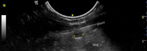
While you have the opportunity, it’s a good idea to have a look at the lungs.

This is a 3mm diameter metastatic lesion in the peripheral lung field. Lesions down to 1mm diameter are detectable so long as they contact the pleura. Mets characteristically have a nummular (round) appearance with little change in surrounding lung. Pulmonary thromboembolic infarcts and pneumonia typically cause marked increase in B lines around the periphery of their hypoechoic lesions.


