Paroxysmal supraventricular tachycardia in an old Bouvier
It’ll be sad if vaccine technology moves on to the point where we don’t need to see cats and dogs at least once a year -I guess that for dogs in the UK it’s hanging on by a perhaps-slightly-tenous Leptospirosis thread at present. My guess is that a lot of owners won’t bring them for routine health checks if no booster required. Anyway, that’s a whole can of worms.
The point is that this case was presented for a booster -whereupon a rather dramatic arrhythmia was apparent. On further questioning it emerged that he had been lethargic recently.
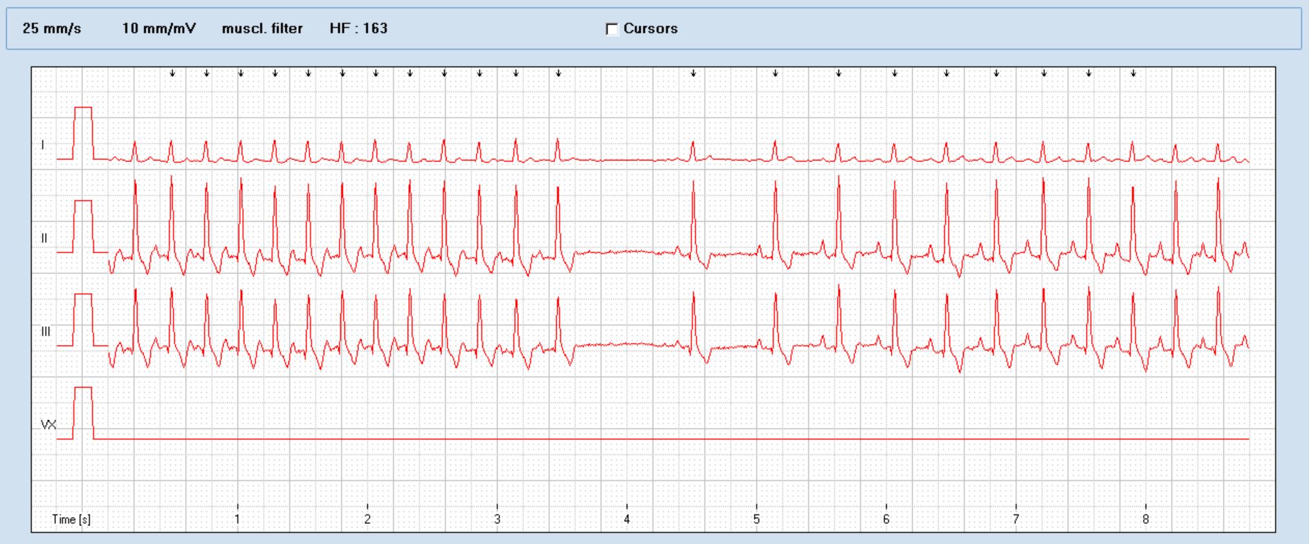
This appears to me to be a paroxysmal narrow-complex (presumed supraventricular) tachycardia. Half way through this section of trace he converts from SVT to sinus tachycardia. Vagal manoeuvres were not effective in achieving conversion.
This is another look at a period of sinus tachycardia at 50mm/s.
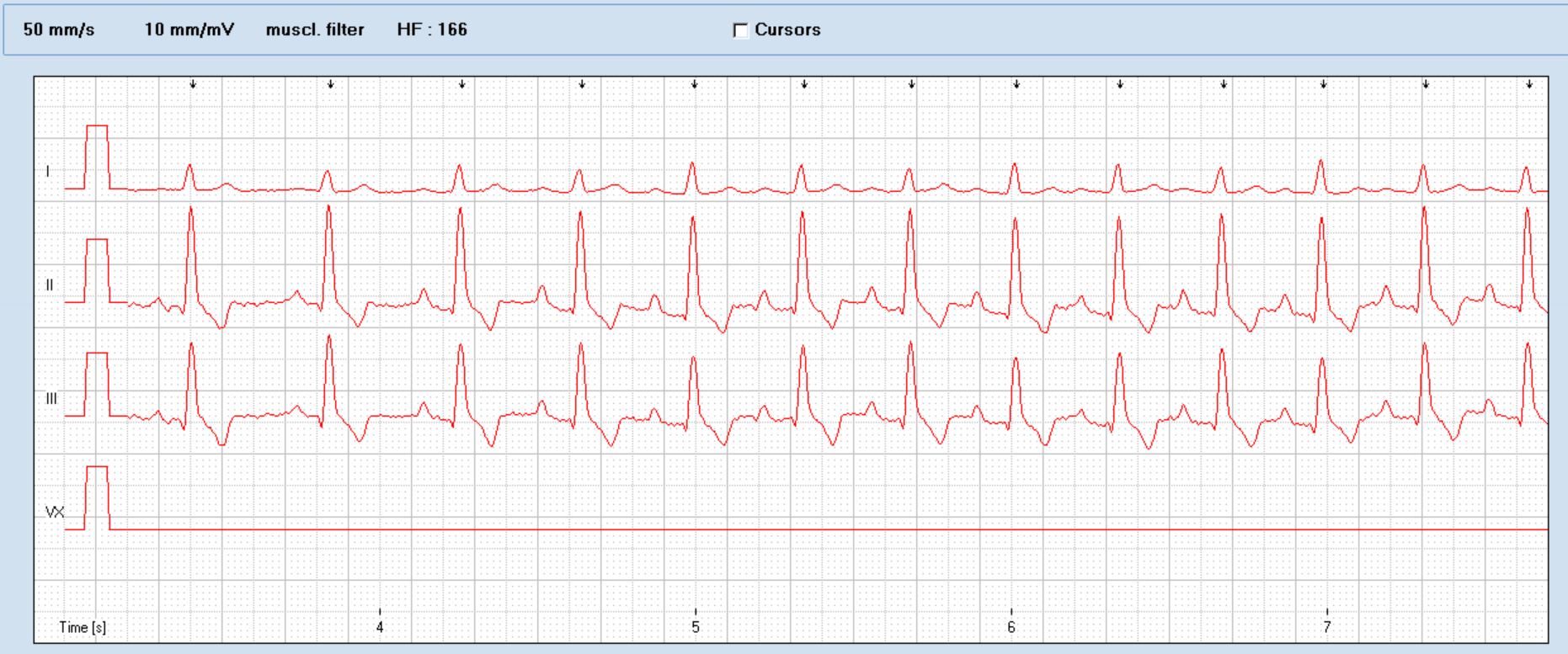
And the SVT, also at 50mm/s
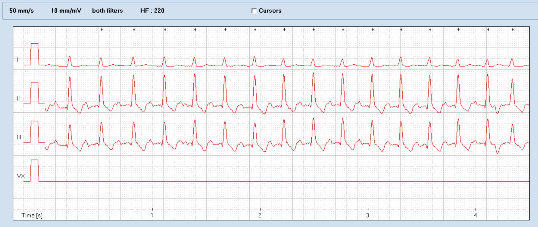
Interesting that the p waves are very similar in both rhythms. It may be that this is a sinus nodal re-entrant tachycardia.
Supraventricular tachycardias in dogs were reviewed by Finster et al.
http://onlinelibrary.wiley.com/doi/10.1111/j.1476-4431.2008.00346.x/abstract
These dogs could broadly be split into those with cardiac pathology and those with non-cardiac conditions.
The present case has no obvious evidence of primary heart disease:
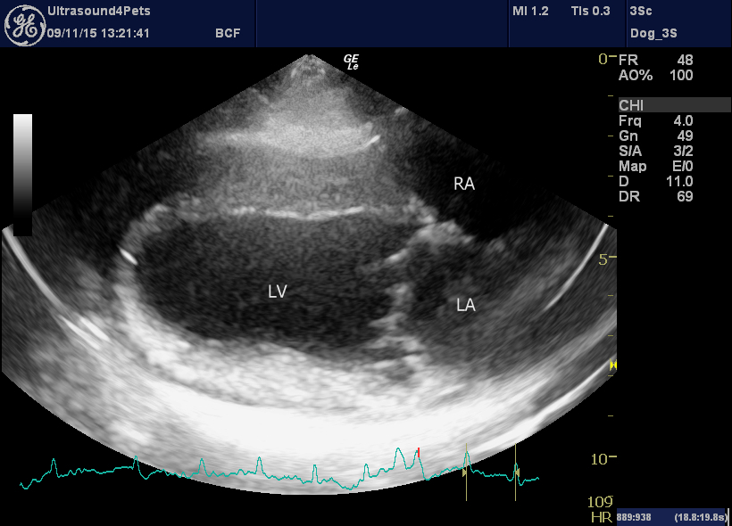
End-diastolic right long-axis 4 chamber view of the heart. All chambers are broadly in normal proportion to each other (the RV is not optimally seen in this frame) and the septae are not bowed.
There was no ultrasonographic evidence of pulmonary oedema and no tachypnoea.
No space here for a full set of echo images but 2D, M mode, spectral and colour Doppler examinations were all unremarkable. LV ejection fraction during sinus rhythm was marginally low at 40% -but this may well be due to tachycardia-induced cardiomyopathy. Other indices of systolic function were within normal limits.
OK, so the non-cardiac dogs in Finster et al.’s series suffered a miscellany of serious conditions from neoplasia to GDV to thromboembolic disease. Routine haematology and biochemistry were unremarkable in our case. On abdominal ultrasound there was just this one lesion in the liver:
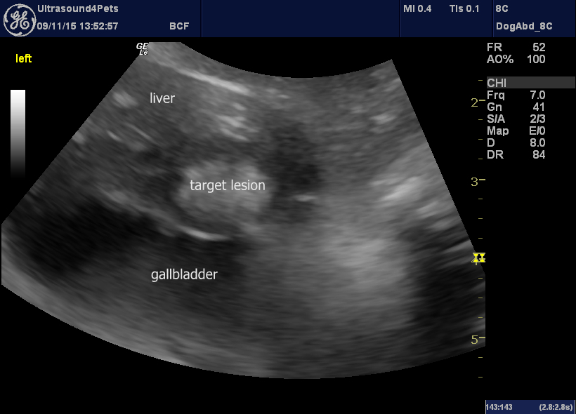
Longitudinal lane view of part of the liver and gallbladder.
The same lesion in transverse plane:
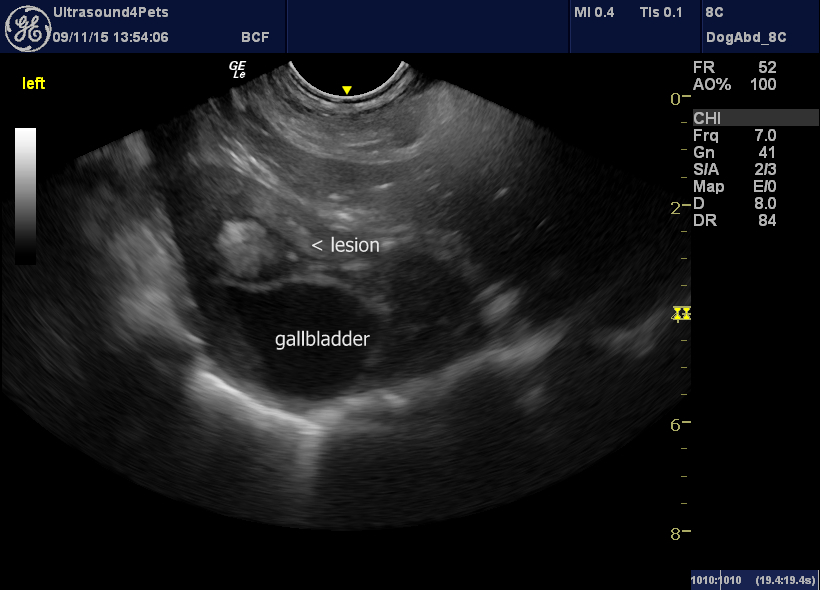
Well, it’s only one lesion -but it is convincingly ‘targetiform’ (jn that it consists of a peripheral hypoechoic zone and a hyperechoic centre) and several publications tell us that hepatic target lesions are ominous.
http://www.ncbi.nlm.nih.gov/pubmed/12088324
http://www.ncbi.nlm.nih.gov/pubmed/22931398
…so perhaps a 66% chance of malignancy even on the presence of this one lesion.
In the event, this poor guy deteriorated rapidly even in the few days as we organised Holter monitoring to establish the pattern of arrhythmia in his home environment and was eventually euthanased. No post mortem was performed.


