Pancreatitis images
Just a few pictures from the last week to illustrate just how common pancreatitis is in both cats and dogs….and how much difference a high-frequency linear probe makes.
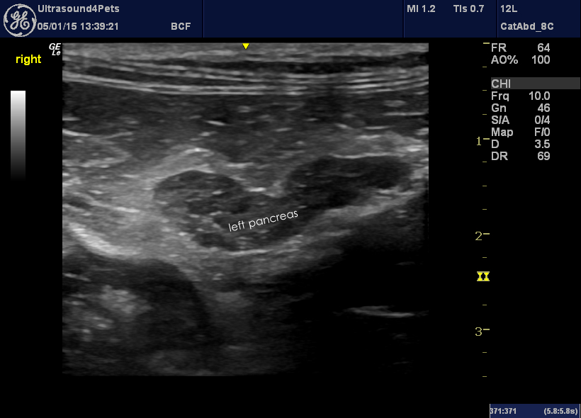
This is the left pancreatic lobe from a cat with chronic recurrent abdominal pain.
The pancreas is ringed by inflamed abdominal fat and the pancreatic parenchyma itself is very heterogeneous.
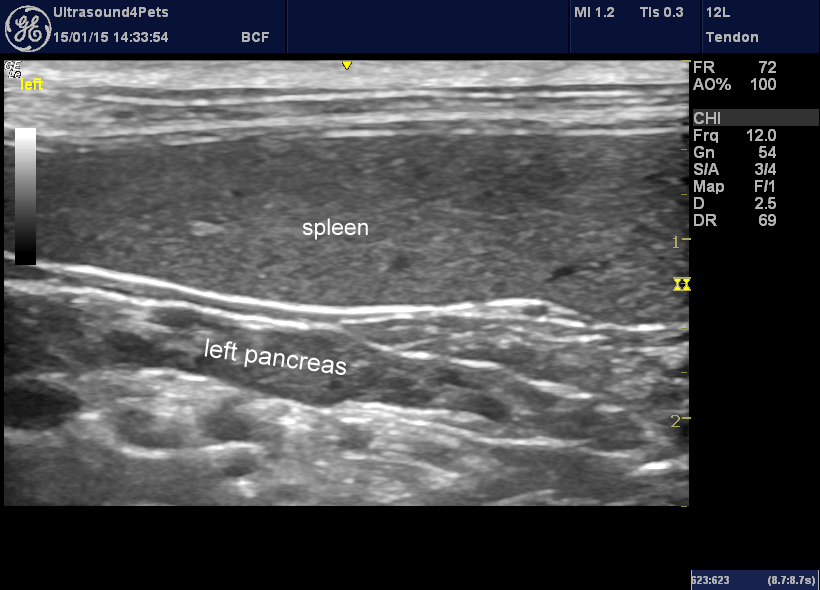
Secondly, an illustration of the fact that the left lobe does lie in an accessible position deep to the spleen in most cats and many dogs. The non-inflamed pancreas is more or less concolorous with the surrounding fat. With a high frequency linear probe and an inflamed pancreas it sticks out like a sore thumb.
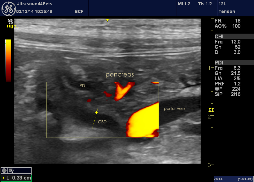
Thirdly details of the common bile duct and pancreatic duct in another cat with acute-on-chronic pancreatitis.
 The common bile duct is seen looping around from cranial (left of the image) and heading for its convergence with the pancreatic duct (PD) at the duodenal papilla. The generally-accepted cut-off for CBD dilation is 4mm in cats. This one is getting close -there may be some constriction in the papilla area resulting from oedema or fibrosis.
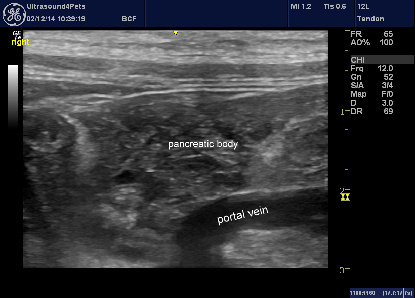
The body of the pancreas itself is again a battlefield of heterogeneous inflammatory change:


