Normal bile acid stimulation test results in two dogs with (presumed congenital) extra-hepatic portosystemic shunts
Although this may not be a big surprise I can’t find any published accounts specifically documenting this phenomenon.
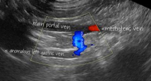
The first case involves a 5 year-old MN Shih-tsu presented on account of haematuria. There was no history of preceding signs suggesting hepatic encephalopathy. No urinary abnormalities were identified on abdominal ultrasound. However, an extra-hepatic shunt vessel was located originating from the gastro-splenic vein, carrying hepatofugal blood flow and following the typical course of an anomalous left gastric vein shunt vessel running cranially and to the left to the diaphragm (presumed either gastrosplenic to azygous or phrenic vein).

longitudinal plane view from the right side showing the main portal vein and its tributaries (under normal circumstances) the mesenteric and gastrosplenic veins. Flow in the gastro-splenic and left gastric veins is hepatofugal.
A bile acid stimulation test (BAST) yielded results basal 24 and post-prandial 4 μmol/l.
On a later date a further BAST showed post-prandial bile acids of 93 μmol/l.
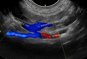
The second case involves a 14 month old FN Coton du Tulear who was examined in the course of a work up for hypoadrencorticism. Prior to signs attributed to adrenal insuffciency she had been asymptomatic. Abdominal sonography revealed an extrahepatic shunt with morphology very similar to the previous case.

A similar right-sided longitudinal plane view of the second case.
Again, BAST results were unremarkable: basal 22 and post-prandial 13 μmol/l.
Again, a repeat at a later date revealed convincingly elevated bile acids: basal 31 and post-prandial 61 μmol/l.
There are numerous potential physiological or artefactual explanations for normal BAST results here. Fully elucidated on the excellent Cornell eclinpath site:
http://eclinpath.com/chemistry/liver/liver-function-tests/bile-acids/
Post-prandial results lower than pre-prandial are not so unusual.
It’s not possible to be definitive about the cause of unexpected findings in our two cases: just to highlight the fact that normal-range BAST results should certainly not be considered to rule out the possibility of congenital EHPSS.
It’s well documented that shunts terminating in the phrenic or azygous veins tend to present later in life:
Vet J. 2014 Mar;199(3):376-81. doi: 10.1016/j.tvjl.2013.11.013. Epub 2013 Nov 23.
Computed tomographic morphology and clinical features of extrahepatic portosystemic shunts in 172 dogs in Japan.
Fukushima K1, Kanemoto H1, Ohno K2, Takahashi M1, Fujiwara R3, Nishimura R3, Tsujimoto H1.
https://www.ncbi.nlm.nih.gov/pubmed/24512983
To quote from the conclusion of that paper ”Spleno-phrenic and spleno-azygos shunts were diagnosed more frequently in older dogs than right gastric-caval and spleno-caval shunts. Dogs with spleno-phrenic shunts had significantly lower fasting ammonia concentrations than those with caval shunts’. The authors hypothesise that shunt fraction was lower or more intermittent in dogs with non-caval shunts….which would fit with our findings in the present cases and adds weight to the theory that their normal initial BAST results weren’t simply artefactual.


