…..more ultrasonography of pneumonia but with added complexity
I’ve blathered on a lot about pneumonia in this blog. It’s still a bit of a revelation to me that, having gone the best part of twenty years diagnosing pneumonia twice a year I now seem to see them twice a week.
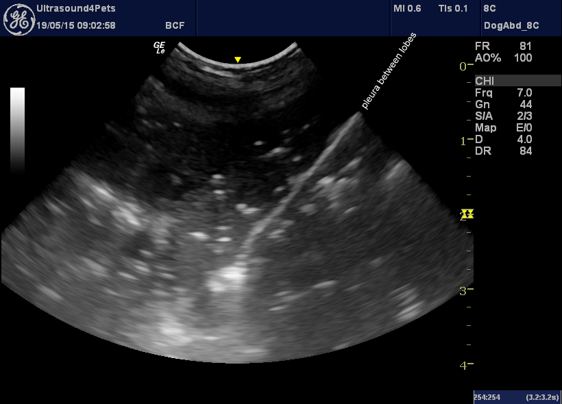
This is a very ill elderly dog with a history of ‘bringing up food’ and now unable to stand and with a rectal temperature of 41C. On survey ultrasonography of chest and abdomen the outstanding finding is this area of abnormal lung in the cranio-ventral chest:
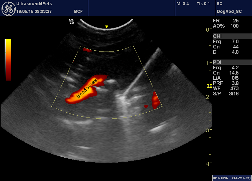
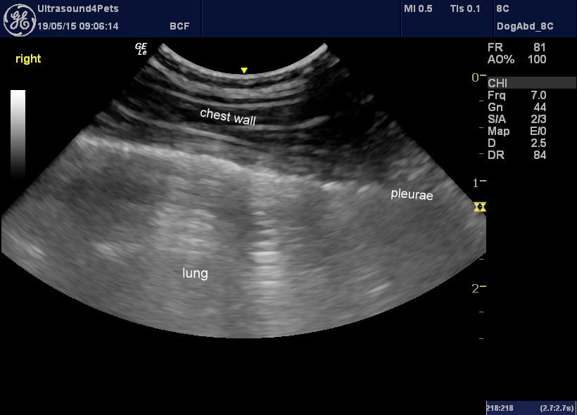
There is consolidation of lung (‘hepatisation’) with a few remnant air pockets showing up as hyperechoic foci. The power Doppler image on the right demonstrates that vascularity persists despite changes in the parenchyma. This is the kind of change which is typical of pneumonia. It’s interesting that the cranioventral lobes are most affected. The dorsal partsof the lung on both sides appear much more normal:
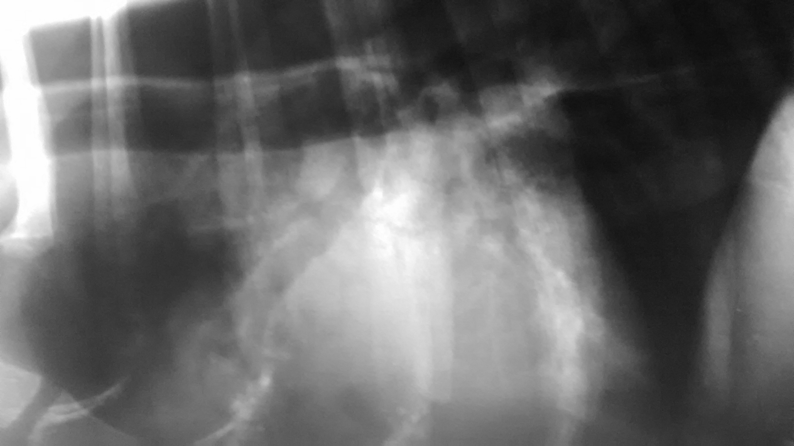
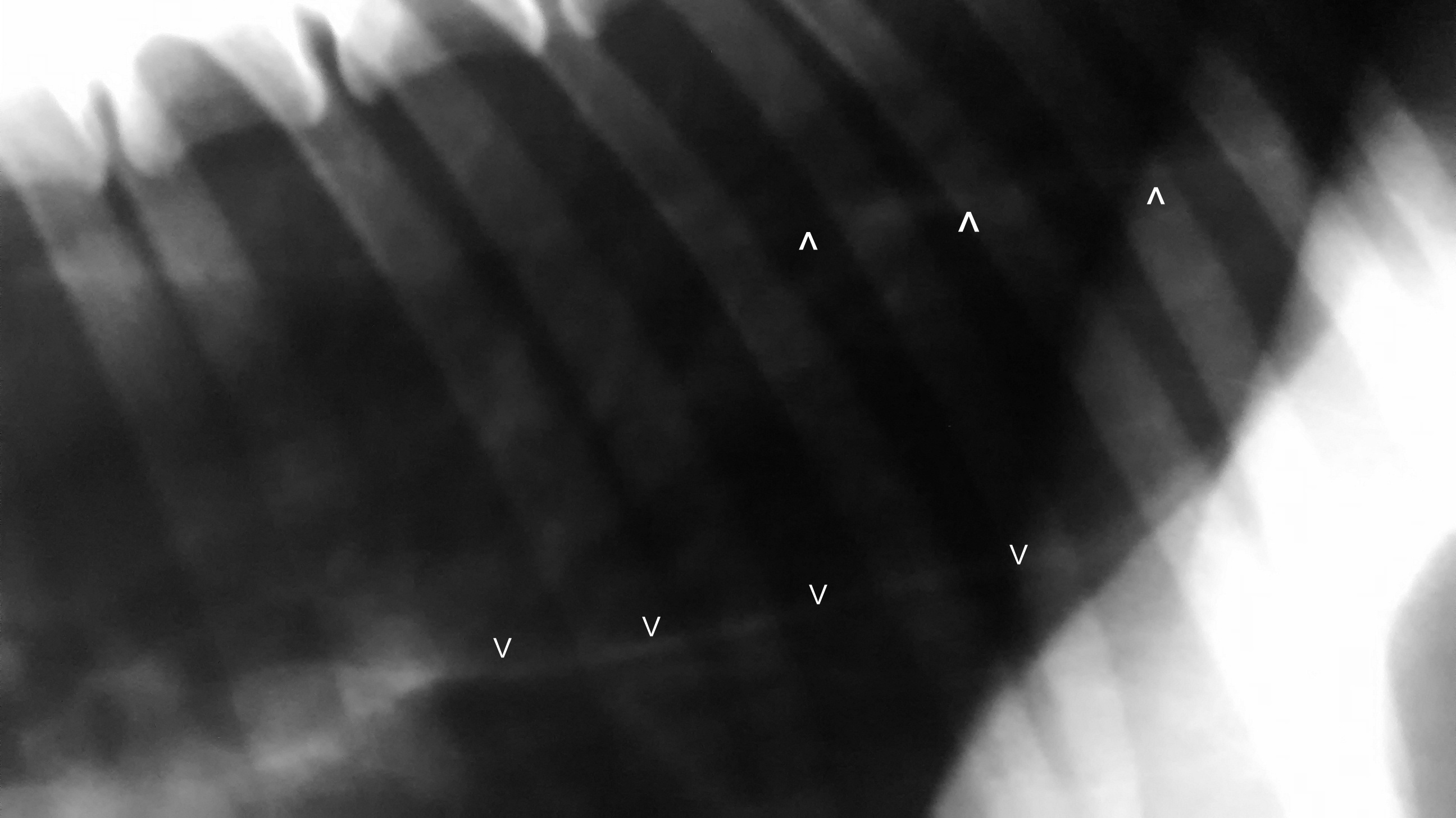
And so to radiography:
Ok, you guessed it -there is megaoesophagus (this is a conscious x-ray) and a typical cranio-ventral distribution of what is presumably inhalation pneumonia.
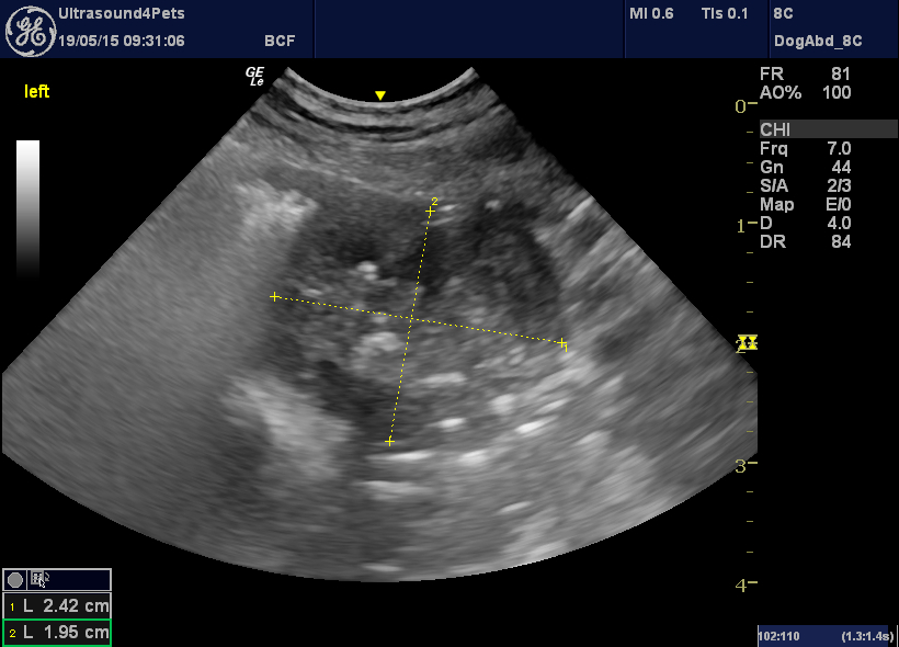
So, why does this 12-year-old dog have a megaoesophagus? This is a closer look at the cranio-ventral thoracic cavity:
This golf-ball-sized mass is likely to be a thymoma. On re-visiting the physical examination, he has a classic fatigable palpebral reflex of myasthenia gravis (his inability to stand, it turns out, may have as much to do with muscle weakness as to the systemic effects of pneumonia). It is highly probable that this is a paraneoplastic myasthenia secondary to the thymoma. The myasthenia-induced megaoesophagus has lead on to the inhalation pneumonia.


