More on the diagnosis of pneumonia using ultrasound
This is a really good recent paper on the subject from human medicine:
Emerg Med J. 2012 Jan;29(1):19-23. doi: 10.1136/emj.2010.101584. Epub 2010 Oct 28.
Lung ultrasound is an accurate diagnostic tool for the diagnosis of pneumonia in the emergency department.
In summary these authors found that, in an A&E setting, the intial chest xray was diagnostic for pneumonia in 18/26 cases whereas ultrasonography was positive in 25/26. They add that ‘The feasibility of ultrasound was 100% and the examination was always performed in less than 5 min’.
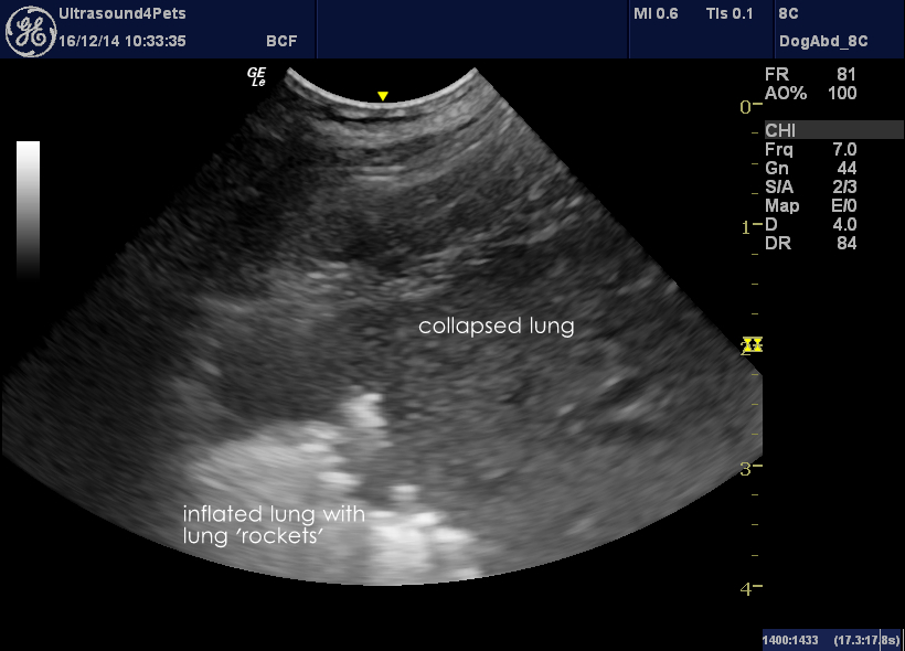
I can confirm from very recent experience that it’s a hell of a lot easier to scan a Great Dane’s chest than to perform radiography. This particular patient presented with a pyrexia of 40.8C. Her chest scans are dramatic:
She has large irregularly delineated areas of consolidated lung on both sides although more extensively on the left. In this video clip the pleura-pleura division between left cranial and left middle lung lobe traverses the image diagnonally. Ultrasound lung rockets (indicative of ‘wet’ lung) arise at the peripheral boundary of the cranial lobe and at the boundary between the lobes. Lung rockets move with the lung during respiratory movements.
[wpvideo tujiUTMS]
One more reason to scan every patient!


