More on feline ureteral calculi and renal infarcts
I’ve written before about the possible link between ureteral calculi and renal infarction in cats. The more of these cats we see the more I suspect that calculi are very common and also that most of them pass through undiagnosed. My hypothesis is that this might explain the number of cats we see with renal cortical hyperechoic wedges.
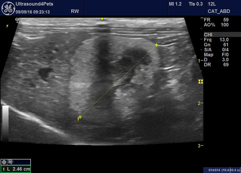
This is a case from last week: acute onset anorexia, malaise, azotaemia.

left kidney short axis view: dilated proximal ureter and 3mm diameter ureteral calculus. There is a hyperechoic, sunken area on the left of the kidney as we see it in this image which would be typical of an infarct -possible from a previous episode of obstruction.
The contralateral kidney has evidence of previous ‘events’: being small and irregular.
Right kidney longitudinal plane view
Doesn’t look promising does it?. One very severely affected, shrunken kidney with presumed chronic changes and the other now obstructed with a huge ureteral calculus. There is urine in the bladder though -so I’m guessing the right side has some residual output.
Now the left kidney 2 days later:

There is a nephrolith in the pelvis but the ureter has relatively normal diameter:
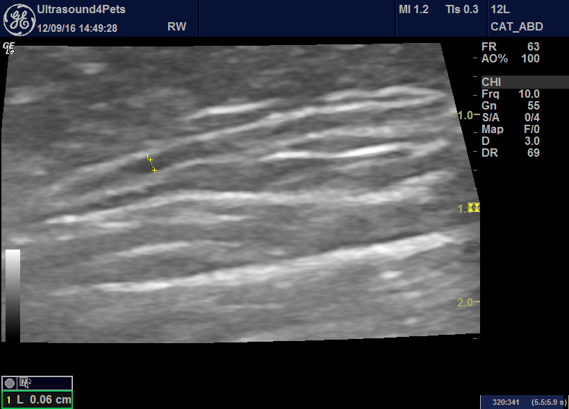
By this time the cat was happily eating again. Amazing what will pass through a ureter!


