….more on chest scanning in abdominal cases
To carry on from the previous post: another case in point although with a less happy outcome.
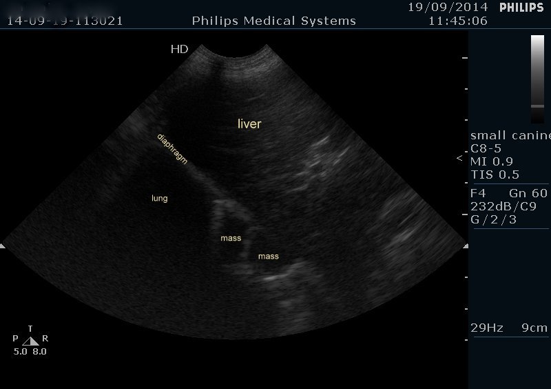
An older Staffie with occasional vomiting. This is a sagittal plane view of the cranial abdomen and diaphragm -again looking from a window just caudal to the xiphisternum. First view in a routine abdominal series.
Already that’s looking ominous. There are multiple soft tissue lesions in the caudal lung field adjoining the diaphragm. Pulmonary mets are often visible on ultrasonography -either through the diaphragm or with intercostal views. They can only be seen if the masses reach the pleura. But in patients with numerous pulmonary mets they usually do.
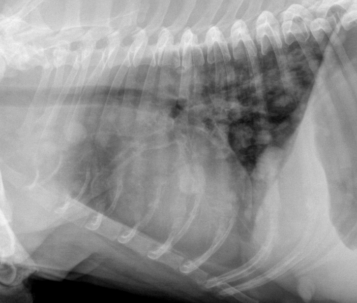
So this is her chest x-ray. Not good.
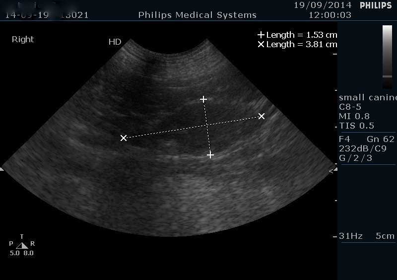
And on completing the abdominal scan she also has a suspect medial iliac (sublumbar) lymph node with an irregular silhouette and heterogenous parenchyma.
Heterogeneous lymph nodes in cats may be caused by inflammatory disease such as IBD or FIP. In dogs, heterogenous lymph nodes carry a higher probability of neoplasia.


