More adventures with shunts
The expansion of veterinary knowledge continues apace. It sometimes feels a challenge just to keep up to date with the state of play even one small area like portosystemic shunts.
The excellent and seminal 2008 edition of ‘Atlas of Small Animal Ultrasonography’ unequivocally states that a portal vein:aorta ratio >0.8 rules out a congenital extrahepatic shunt. For a long time this has been a given.
Now we have…
Computed tomographic morphology and clinical features of extrahepatic portosystemic shunts in 172 dogs inJapan.
….which contains the revelation that PV:aorta ratios were as follows for diiferent shunt types
Spleno-phrenic 0.37-1.31
Spleno-azygous 0-1.3
R gastric-caval 0-0.75
Spleno-caval 0-1.03
And furthermore phrenic (64/172 cases) and azygous (38/172) were the commonest types.
Back to the drawing board then!
This week’s shunt case: a spleno-phrenic PSS in a kitten:
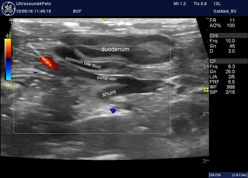
Longitudinal plane view from R flank behind last rib looking at the porta hepatis
This is the standard porta hepatis view which is key in many cat abdomen examinations. The portal vein is narrower than the mesenteric vein/splenic vein confluence caudal to it: which always suggests the likelihood that blood is being diverted down a shunt through the spleno-left gastric vein.
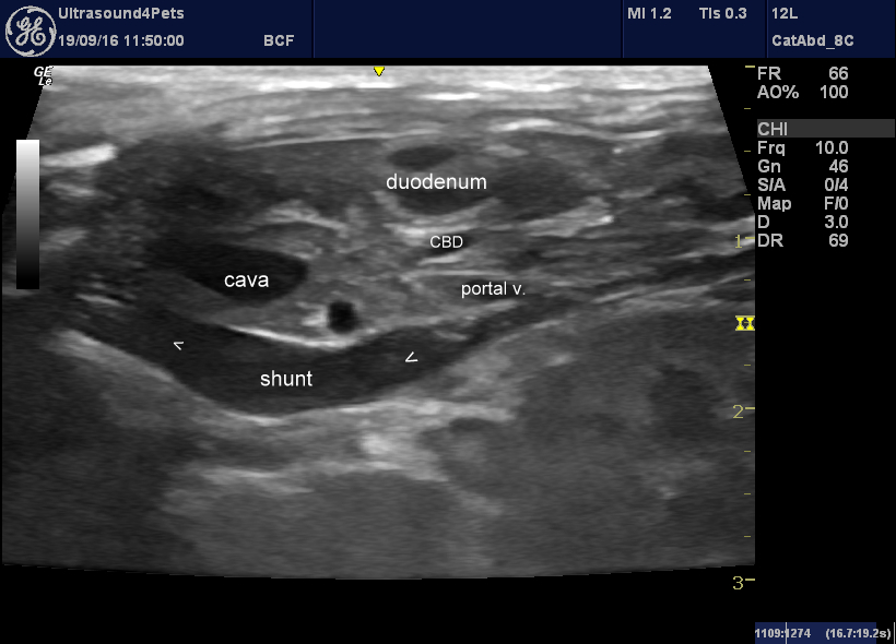
same view, subtly different angle to follow the shunt
And indeed the left gastric vein does lead into a large abnormal vessel coursing across the lesser curvature of the stomach to the left of the portal vein.
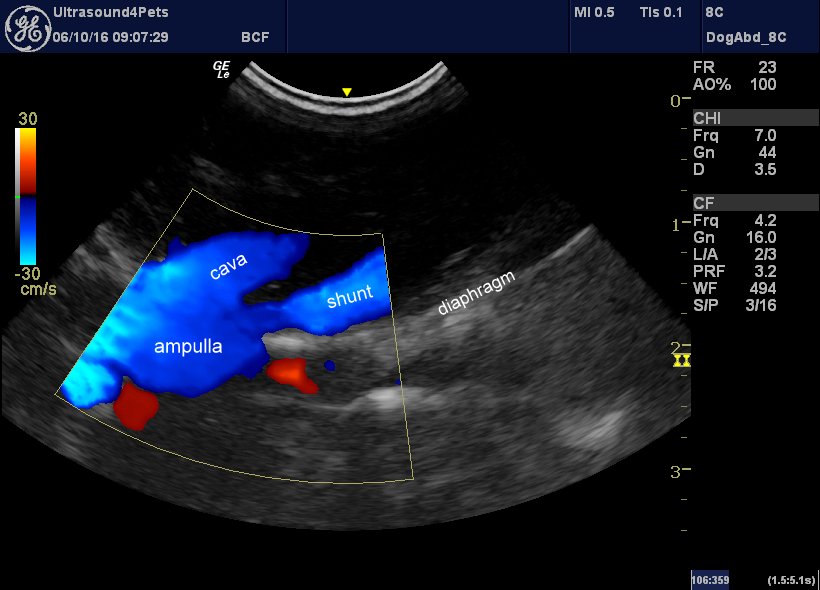
Oblique view at the diaphragm from right side showing confluence of shunt and cava at the level of the diaphragm with a large ampulla at this point
This is typical of left phrenic shunts: the shunt reaches the diaphragm joining the phrenic vein and then tracks across the diaphragm to join the cava; often with a dilated ampulla.
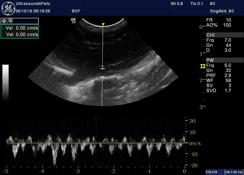
With PW Doppler shunt flow fluctuates markedly with the cardiac cycle (suggesting continuity with the cava).
At surgery, from the right side after medal reflection of the duodenum:
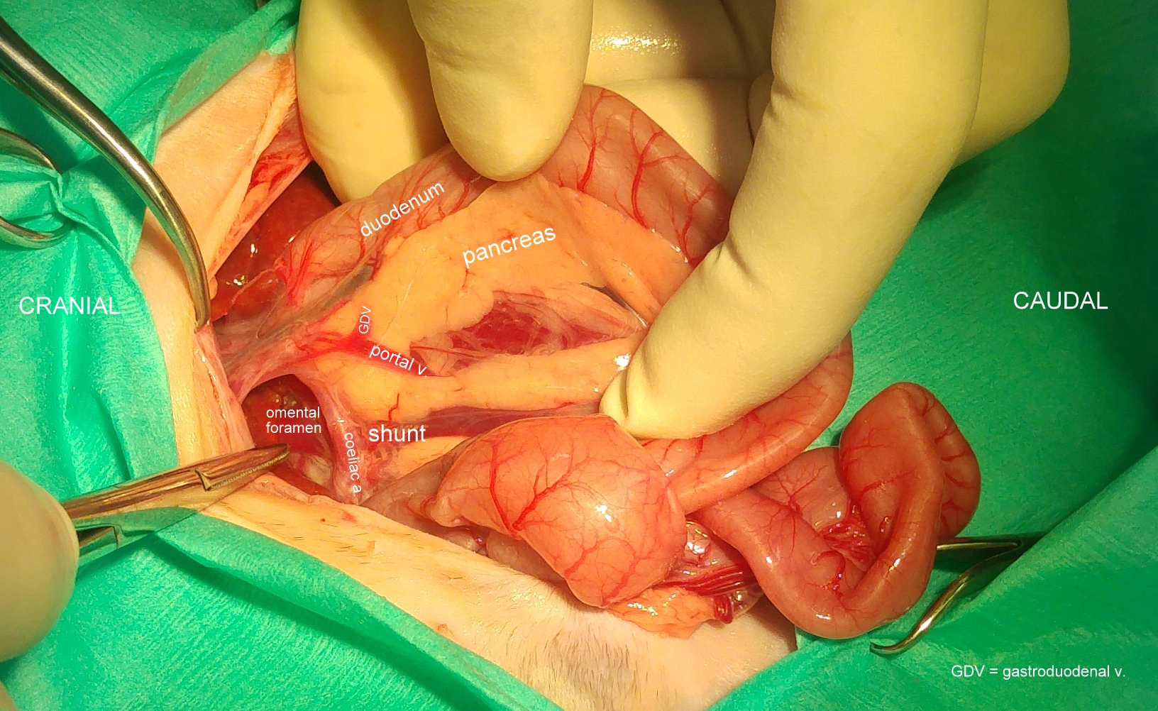
That’s pretty much the view we were seeing in the first sonographic image above. I haven’t labelled the bile duct but you can see it between duodenum and portal vein.
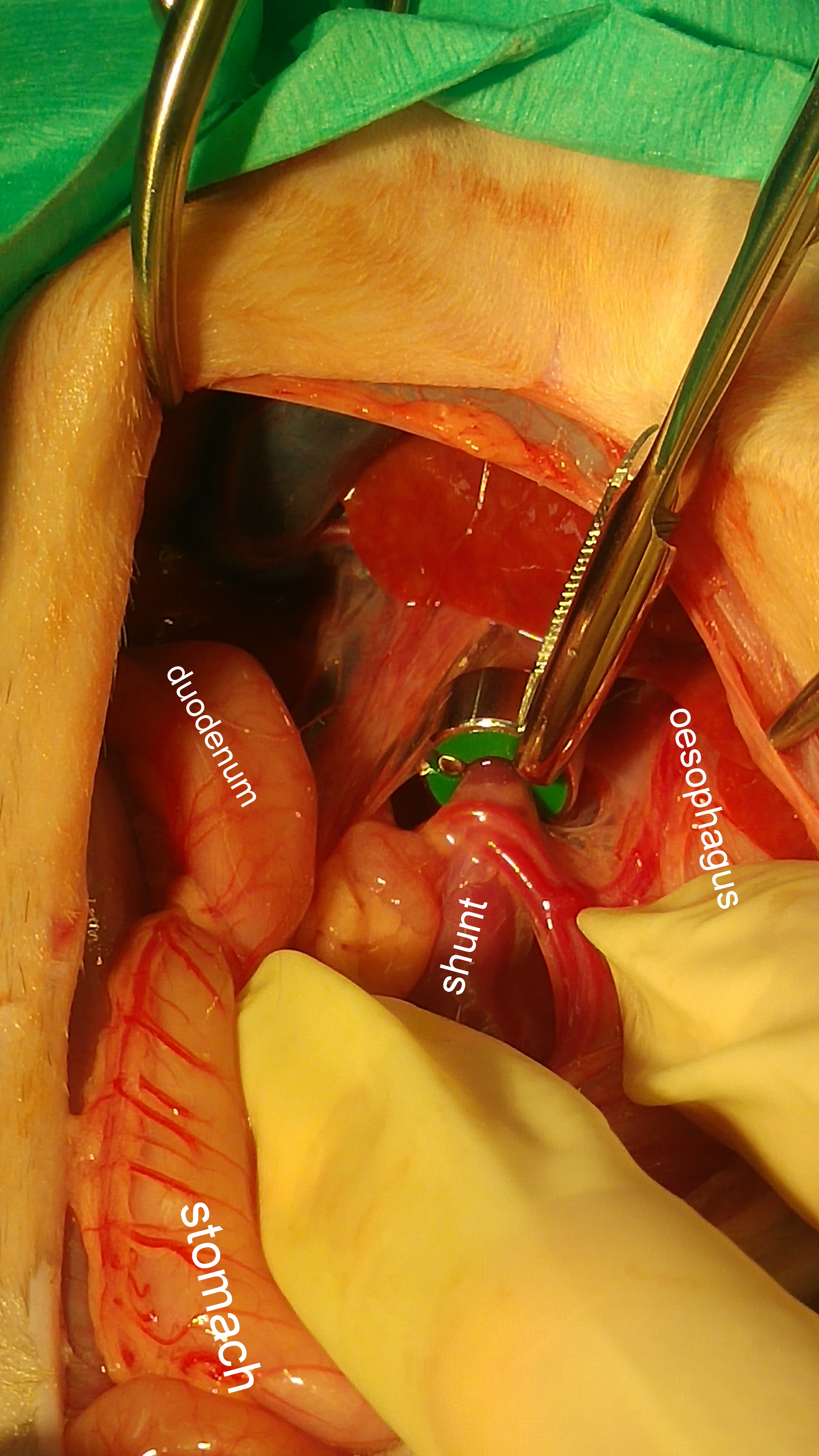
And the shunt at the moment of its demise by ameroid constriction-fingers crossed.


