Lung ultrasound protocol
A little salutary tale from my week.
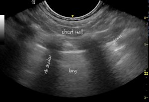
This little dog was presented with a history of cough and, primarily, I was asked to perform an echo. My philosophy is that it’s good routine to do a full thoracic exam along with any echo and also to check the abdomen.
This I duly did. Nothing abnormal to be seen.

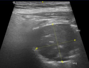
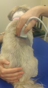
Writing up her report and having another read of her history I realised that she’d had a prior chest xray and that a suspect opacity had been noted in the dorsal lung field. So, back to the patient and with a clip up to the dorsal midline this, rather obvious, lesion is now visible.

This is the view required to see it:

I’ve come across lung lesions like that before. In particular those affecting the accessory lobe of the right lung may only be accessible from the extreme dorsal chest wall.
Difficult isn’t it! Nobody likes their dog to come back from the vets completely bald but sometimes it’s necessary. Either that or really slather with spirit for the dorsal bits which will be visible in the park.


Hi! thanks very much for share all this cases. Did you were alow to PAF it? it could be a pneumonic focus?
Thank you very much.
Thanks Joanna,
We didn’t take any biopsies in the end -she is about 13 years old and the owners decided they didn’t want to do any further testing.
If it was pneumonia then I would expect that the margins of the consolidated hypoechoic area would be serrated, there would be residual gas bubbles within the lesion and the surrounding lung would have lots of B lines indicating oedema (in the broadest sense; not cardiogenic obviously).
…like this https://youtu.be/gymnJNG5df8
…although that one is a cat