Lung ultrasonography in canine Angiostrongylus lungworm infestation
The last cases of Angiostrongylosis we saw in the frozen north (West Yorkshire) were following the very mild winters of the mid-noughties. So it may be no coincidence that a resurgence happens now.
This is a dog from earlier in the week with positive A. vasorum serology. The pulmonary hypertension is fairly standard:
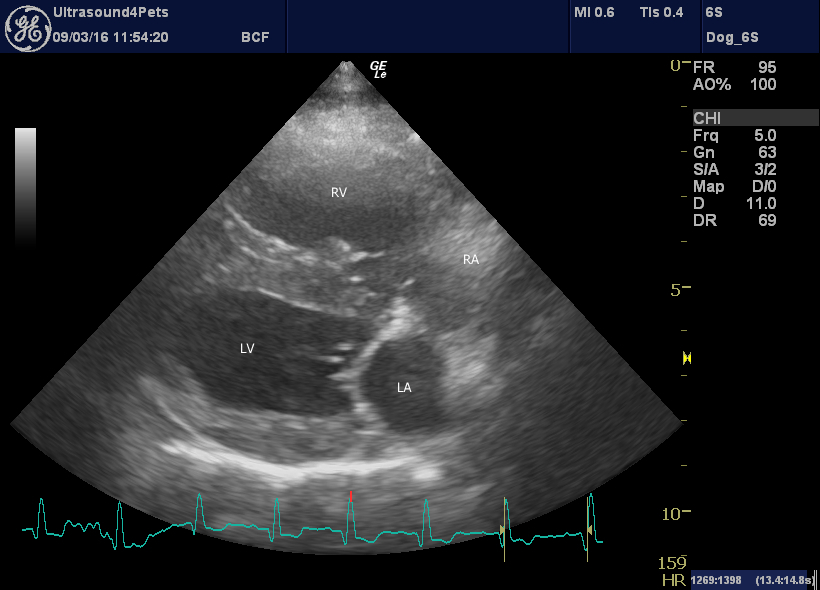
Right long axis 4-chamber view: there is pronounced right ventricular dilation, systolic flattening of the ventricular septum to the left.
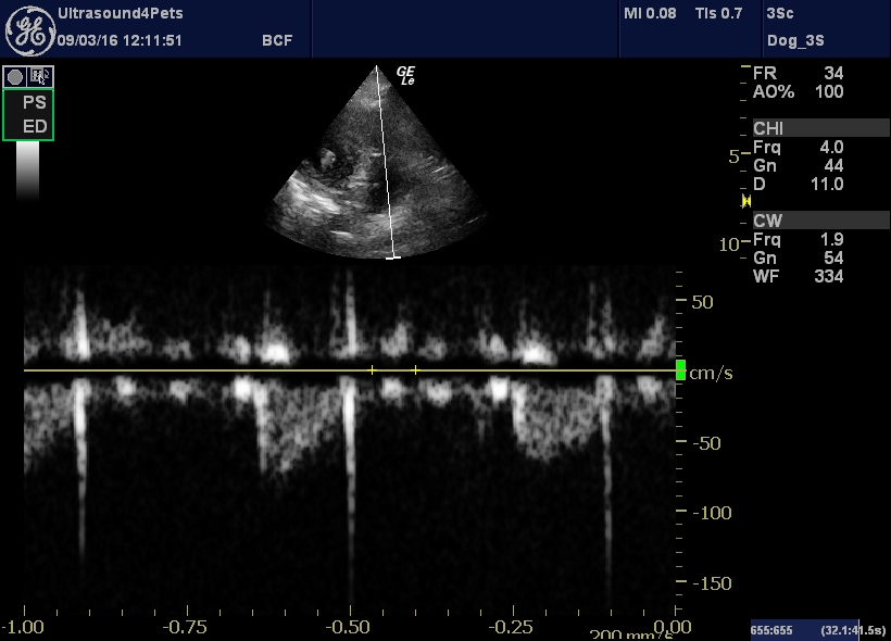
left cranial view of the pulmonary artery with CW Doppler. The flow profile is markedly asymmetrical with an acceleration time:deceleration time ratio 0.2
This pulmonary artery AT:DT is much lower even than reported by Tidholm et al. for dogs with pulmonary hypertension secondary to mitral valve disease.
http://www.ncbi.nlm.nih.gov/pubmed/26365438
We were unable to document any significant tricuspid regurgitation.
The most interesting thing for me was the appearance of the lungs:
This looks identical to cardiogenic pulmonary oedema to me: increased ‘B lines’ and fuzzy lung margins.
This is cardiogenic pulmonary oedema for comparison:
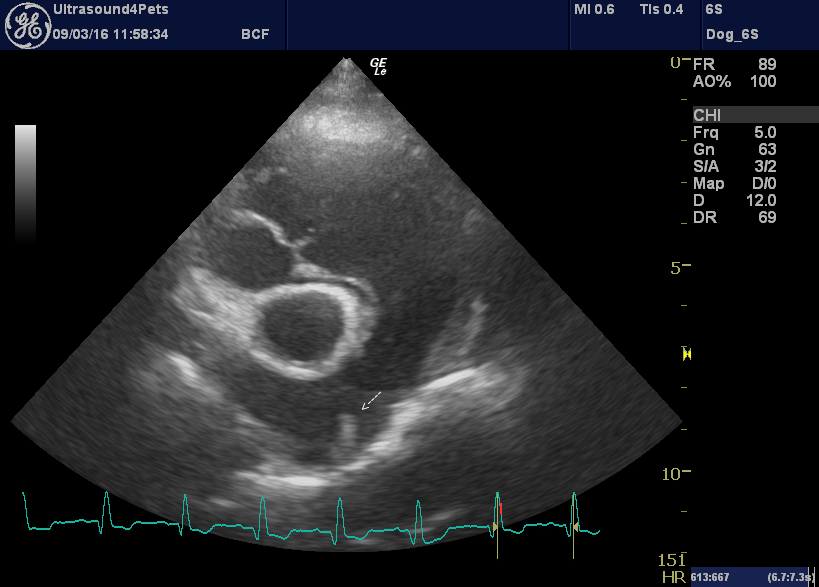
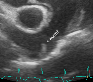
Finally, a right transverse image of the pulmonary artery. The main pulmonary artery is very dilated. Hard to be sure but there is an abnormal structure at the bifurcation which could even be a worm.