Intravesical ureterocoele in an adult Labrador
Advanced warning that this article includes one of the most amazing ultrasound images I’ve seen in some time.
The patient is an 8 y.o. entire female Labrador presenting with acute malaise and pyrexa 40.5C. I’m a firm believer in the maxim that any dog with a rectal temperature >39.5C deserves survey ultrasonography of chest and abdomen.
First 2 seconds of the abdominal exam and we have an interesting finding mid-abdomen:
longitudinal view of the dorsal mid-abdomen: an entire bitch…..pyrexic….large loops of fluid-filled viscus in the abdomen….surely……
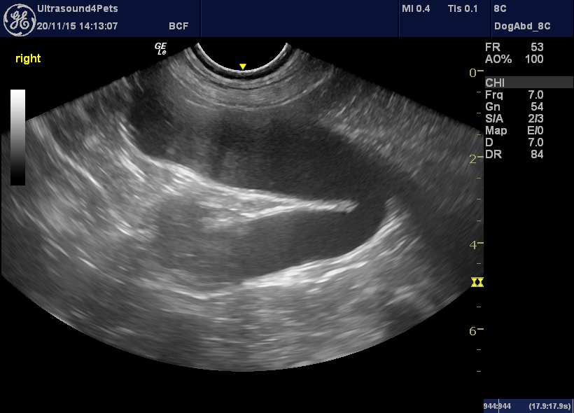
Well, diarrhoea in the colon is an important differential in dogs with putative pyometra. But, the wall of this structure doesn’t immediately strike me as likely gastrointestinal tract -the layering is not typical. On the other hand it’s a bit thin for uterus too. Look what happens now:
Wow! waves of muscle contraction. Well, canine uterine horns don’t normally do that -whether pyometra, mucometra/hydrometra or whatever.
Anyway, onwards with the examination. So this is the bladder area as seen from the ventral mid-line:
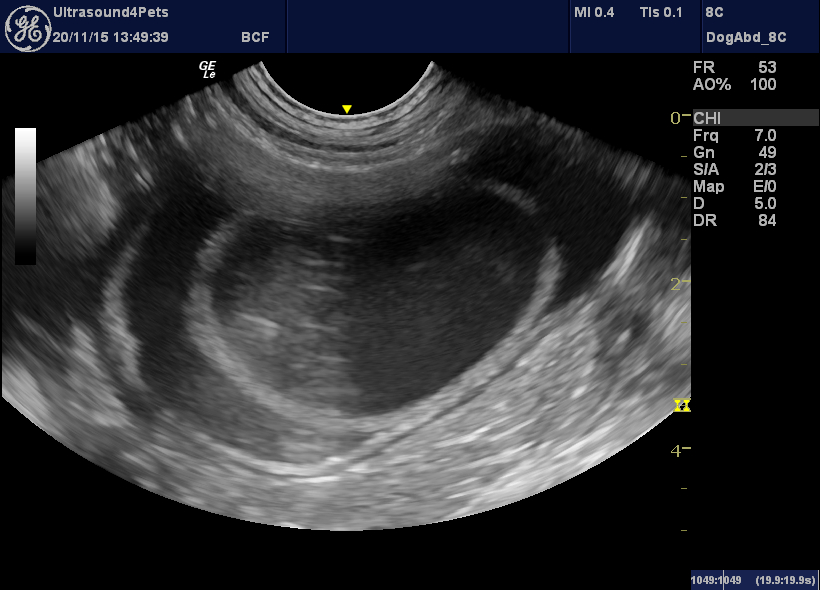
Hmmm, that was a little unexpected!
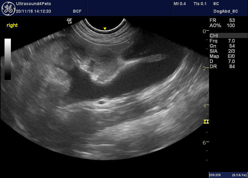
On further exploration the inner compartment communicates with the fluid-filled structure cranial to the bladder:
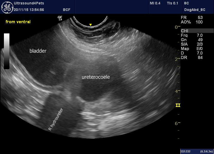
… and that structure is revealed to be the right ureter. There is a unilateral hydroureter with pyelectasia (dilation of the renal pelvis).
The ‘bladder within the bladder’ is a ballooning of the intramural section of the distal ureter into the bladder lumen as an intravesical ureterocoele.
I presume this to be congenital. A definite cause of pyrexia was not identified although she responded promptly to antibiotics and I guess may have had a UTI.


