Intra-operative ultrasound: utility in intrahepatic shunt attenuation in dogs
The use of intra-operative ultrasound in canine shunt surgery has been described previously:
J Am Vet Med Assoc 2003 Apr 15;222(8):1086-92, 1077
Intraoperative ultrasonography of the portal vein during attenuation of intrahepatic portocaval shunts in dogs
Viktor Szatmári 1, Frederik J van Sluijs, Jan Rothuizen, George Voorhout
https://avmajournals.avma.org/view/journals/javma/222/8/javma.2003.222.1086.xml
The authors of that study interrogated the main extrahepatic portal vein with PW Doppler in order to assess the severity of portal hypertension after shunt ligation.
In a recent case, a young Deerhound with a left divisional intrahepatic shunt, due to financial constraints we were unable to perform pre-op CT angiography. Instead, we used intra-operative ultrasound (with sterile probe cover and sterile gel) to confirm that we had encircled the correct vessel with a ligature prior to placing a cellophane band.
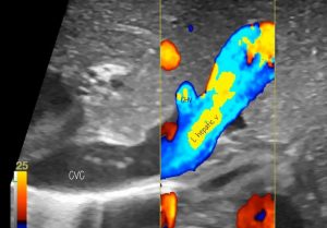
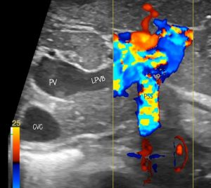
In all of these images the linear probe was inserted between the cranial face of the liver and diaphragm to examine the major vessels. The shunt vessel was encircled at a post-hepatic site (between liver and diaphragm).

Intrahepatic ultrasonography of intrahepatic vessels: transverse plane with L liver on R of image. CVC caudal vena cava, CHV central hepatic vein. The L hepatic vein is very dilated

Intrahepatic vessels: same view slightly adjusted to optimise for portal vein. PV portal vein, LPVB left portal vein branch, PSS portosystemic shunt, CVC caudal vena cava
In video, you can see how these relate to each other:
The shunt enters the distal part of the left hepatic vein. The configuration shown in figure 4A and B of the indispensible:
Vet Radiol Ultrasound 2020 Sep;61(5):519-530.
Canine intrahepatic portosystemic shunt insertion into the systemic circulation is commonly through primary hepatic veins as assessed with CT angiography
Mark J Plested 1, Allison L Zwingenberger 2, Daniel J Brockman 1, Silke Hecht 3, Scott Secrest 4, William T N Culp 3, Randi Drees
https://onlinelibrary.wiley.com/doi/10.1111/vru.12892
When we temporarily tightened the ligature, flow in the shunt was attenuated…which was encouraging:
The patient continues to do well at the time of writing a couple of months later.
This is not going to become the gold standard technique. Sonography of shunts is highly operator-dependent and lacks the objectivity of angiographic techniques. All studies show lower accuracy with ultrasound. However, it’s a handy budget option.


