Hypercalcaemia in a dog
With a really marked hypercalcaemia in a seven-year-old dog one always hopes for primary hyperparathyroidism with a nice parathyroid nodule to remove.
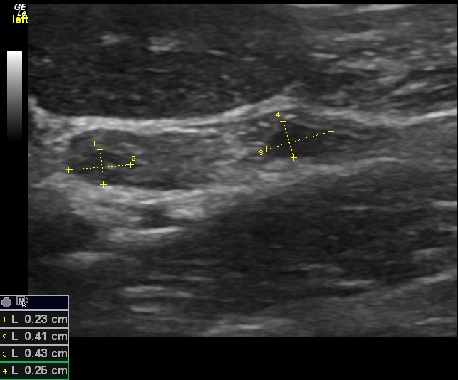
So, here is the left thyroid (linear probe, just love it!)
…and those look like sizeable parathyroids right?
Well, probably not actually. Parathyroids in perfectly normal dogs can be up to 7.5mm long and 3.3mm wide (Liles et al. Vet Radiology and Ultrasound 2010). Not only that, it’s also pretty much impossible to be completely sure of the identity of hypoechoic structures within the thyroid -in the same study 9/35 structures identified as candidate parathyroids actually turned out to be lobular thyroid tissue.
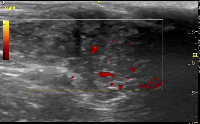
We went on to look at the entire chest, abdomen and anal glands. Sadly, this guy has serious issues. The left anal gland contains a vascular soft tissue mass about 2cm diameter.
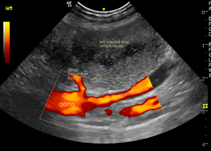
And, even worse, the ipselateral medial iliac (sublumbar lymph node) is grossly enlarged with an irregular silhouette and heterogeneous echotexture. This spread often occurs early in the course of disease in dogs -it’s not so surprising that the metastatic lesion is large in relation to the primary.
A core biopsy from this lymph node confirms a likely adenocarcinoma consistent with anal gland origin.
That’s not good. However, there are treatment options. Chest radiographs were clear. There are two small case series of dogs in which anal gland resection and iliac lymphadenectomy were performed. Mean survival was 2-3 years. The hypercalcaemia typically resolves within 24 hours of surgery. So, there is hope…..watch this space.


Latest update: the primary mass and the iliac nodes have been removed. Hypercalcaemia did indeed return to normal within 24 hours and the patient is currently well.