Hepatic vein flow in right heart failure and in normal dogs and cats
I have heard it suggested in cardiological circles that alternating bidirectional hepatic vein flow is an indicator of right heart congestive failure. But it’s not that straightfoward.
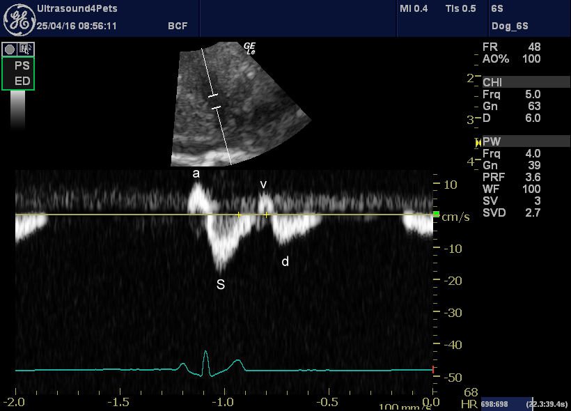
Normal dogs also have not one but two reversed phase in their hepatic vein flow:
As was described originally by Szatmari et al. some years ago:
http://www.ncbi.nlm.nih.gov/pubmed/11327368
The reversed ‘a’ wave corresponds to atrial contraction
The forward ‘s’ wave corresponds to right atrial filling during ventricular systole
The reversed ‘v’ wave corresponds to right atrial overfilling at the end of systole
the forward ‘d’ wave occurs during ventricular diastole as blood flows through the tricuspid valve
The normal flow pattern can thus be described as tetrainflectional:

PW Doppler interrogation of right hepatic vein flow in a normal dog
‘right heart failure’ actually encompasses a variety of conditions with differing (and complex!) effects on hepatic vein flow.
A: Restrictive cardiomyopathy. Systolic (S) forward flow velocity is smaller than diastolic (D) forward flow velocity. Inspiratory diastolic flow reversal is larger than expiratory diastolic flow reversal.
B: Constrictive pericarditis flow pattern is similar to that of restriction except that expiatory diastolic flow reversal is larger than inspitatory diastolic reversal.
C: Pulmonary hypertension. Diastolic flow reversal does not change much with respiration.
D: Severe tricuspid regurgitation causes late systolic flow reversal (if really severe then the whole s wave may be reversed and amalgamated into one a-s-v wave).
All clear then! I’ll try to save some good examples in the next few weeks.


[…] Hepatic vein flow in right heart failure and in normal dogs and cats […]