Grass seed foreign bodies: sonography and ultrasound-guided retrieval
Grass seed (and other vegetation) foreign bodies are a potential nightmare. We all know this. Sadly, it seems that neither CT nor MRI are reliable modalities for locating them in our patients. Obviously there may be clues from advanced imaging findings but actual visualisation of the foreign body itself isn’t consistent.
In a series of 40 cases subjected to CT the actual grass awn itself was only seen in 10%.
Vet Radiol Ultrasound. 2008 May-Jun;49(3):249-55.
Radiographic, computed tomographic, and ultrasonographic findings with migrating intrathoracic grass awns in dogs and cats.
Schultz RM1, Zwingenberger A.
https://onlinelibrary.wiley.com/doi/abs/10.1111/j.1740-8261.2008.00360.x
Sonography is a slightly different proposition in that its diagnostic efficiency is very much operator-dependent and effort-dependent. The longer you look the more you find.
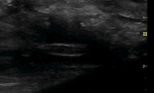
This is a grass seed embedded in the quadriceps of a dog:

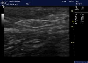
Looks convincing enough doesn’t it? However, it’s not so simple when you have a whole dog to explore. Note that there is no appreciable shadowing. Now look at this from the same dog:

That’s what I would call a ‘pseudo grass seed’. That spindle shape is a small compartment between fascial planes in the subcutis. The differences from the real article are subtle. The genuine grass awn has sharper and more regular margins on a very fine scale. In real time the lines of the husk of the genuine awn finish abruptly and cannot be traced into continuing lines delineating anatomical structures.
Retrieval of grass seeds can be facilitated enormously with ultrasound. This is something we have learned from the excellent paper by Birettoni et al.:
Acta Vet Scand. 2017; 59: 12.
Preoperative and intraoperative ultrasound aids removal of migrating plant material causing iliopsoas myositis via ventral midline laparotomy: a study of 22 dogs
Francesco Birettoni,#1 Domenico Caivano,#1 Mark Rishniw,2,3 Giulia Moretti,1 Francesco Porciello,corresponding author1 Maria Elena Giorgi,1 Alberto Crovace,1 Erika Bianchini,1 and Antonello Bufalari1
https://www.ncbi.nlm.nih.gov/pmc/articles/PMC5310010/
Which was brought to our attention by the equally excellent Valentina and Ron at Ashleigh Vets in Knaresborough.
Rather than open up the entire area with conventional surgery, a good technique is to place a needle under ultrasound guidance through the skin and down to the FB. Make a small scalpel incision through the skin, fascia, muscle sheath and any other relatively tough structures using the needle as a guide. Through this opening a pair of forceps can be passed in order to grasp the end of the seed and withdraw it.


