Gastrointestinal foreign bodies
Interesting cases from this week, happily doing well at present.
The first, a cat with acute vomiting and profound malaise:
The first two seconds of the examination. Don’t want to jump to any conclusions but a stomach dilated to that extent raises the prospect of mechanical or functional GI obstruction.
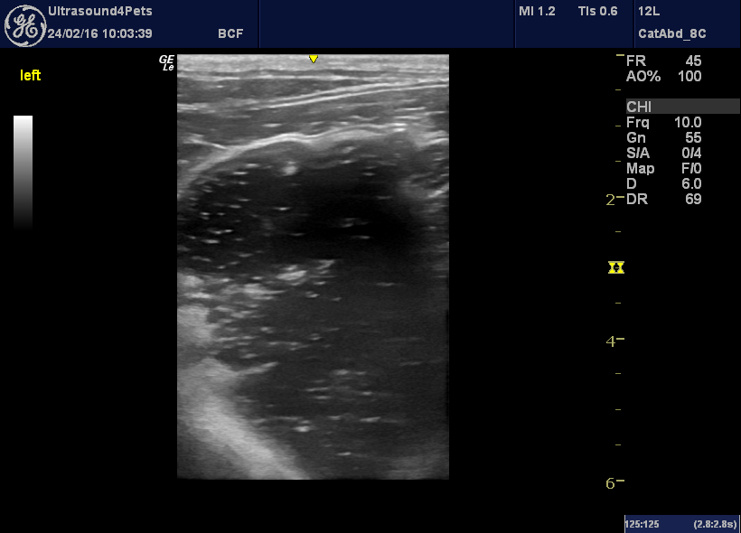
Sagittal plane view of the cranial abdomen: the stomach is massively dilated with liquid content
And there’s some ‘stuff’ in the lumen:
That kind of thing is generally to be regarded with scepticism. A lot of perfectly non-obstructed animals walk around with bits of grass, sweet wrappers, hair and the like in their stomachs.
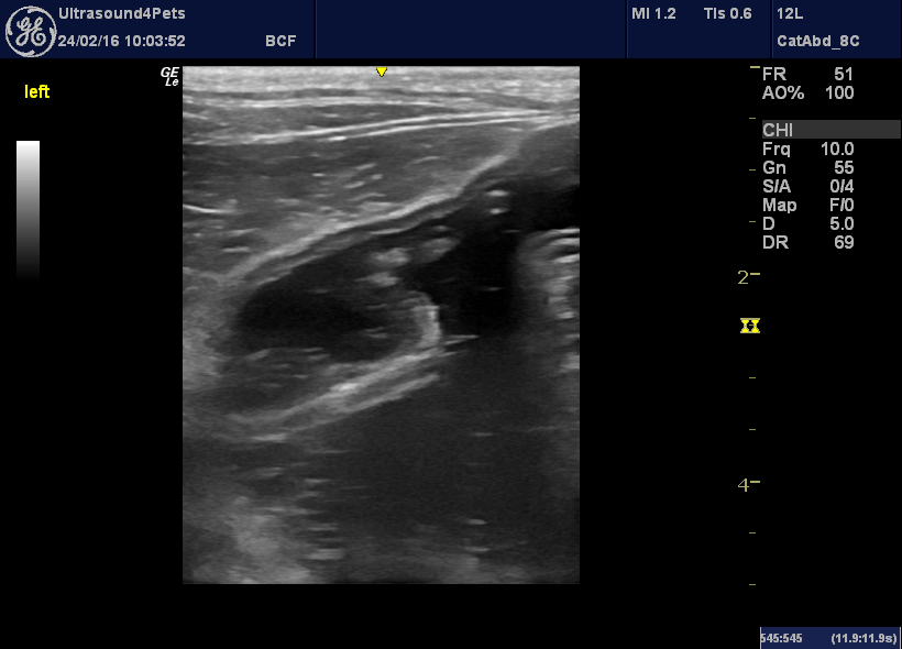
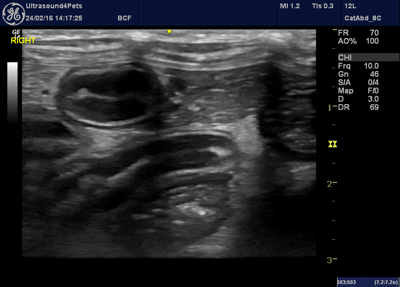
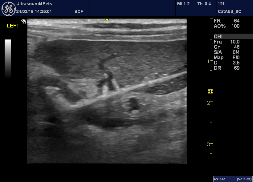
However, here we have the duodenum:
frontal/dorsal plane view of the duodenum from the right sublumbar area
Now I would say that is convincingly plicated (rather than just corrugated). There are real folds rather than crinkles. And there’s a linear thing running through the lumen.
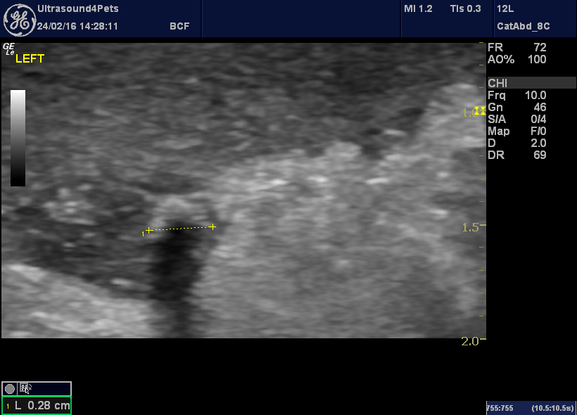
A slightly more detailed view of the linear foreign body. This is the characteristic appearance of the stringy elastic stuff which they use to tie up joints of meat. Thick, bright, hyperechoic lines outside and a thinner central line.
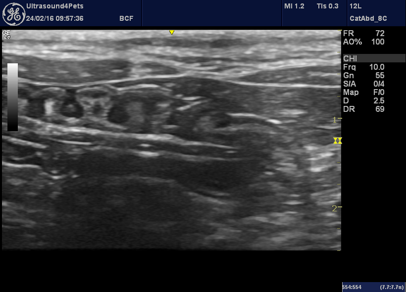
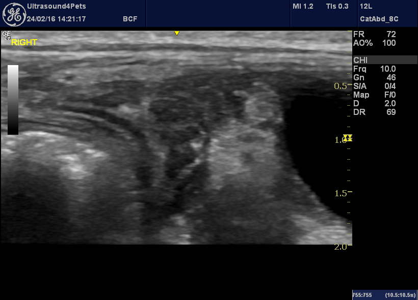
Next up, Yorkiepoo: pyrexic, painful abdomen 48 hrs, positive cPLI.
oblique plane view from right sublumbar area showing descending duodenum in short-axis top left and the right pancreas to the right of it in the image
Any takers for pancreatitis? Looks a bit underwhelming. There was a bit of hyperechoic change in the surrounding fat but…
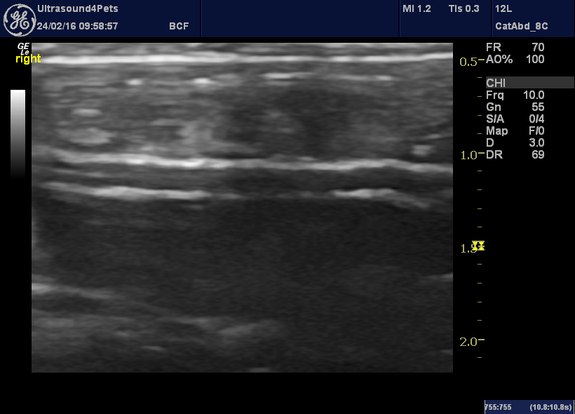
Longitudinal plane view of caudal abdomen with bladder to the right of the image
….the abdominal fat in the caudal right abdomen looks just as hyperechoic.
And on the left….
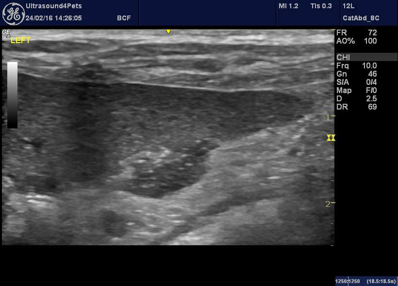
Longitudinal plane from left flank with spleen in the near field
…now were talking! The fat is also hyperechoic but there is also a discrete pocket of turbid fluid deep to the spleen.
same view, slightly different angle
Whoah! and it’s not an artefact. Rotate the probe 90 degrees:
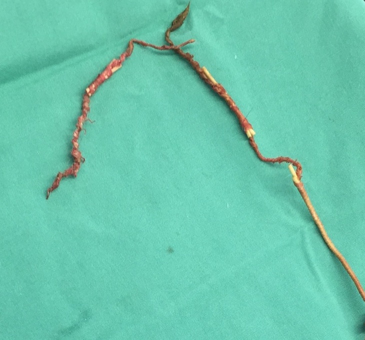
It really is a rigid, linear FB which is exiting that piece of gut (it’s the left colon) on the left of the image and glancing along the underside of the spleen.
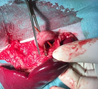
Cocktail/kebab stick through the colon. Ouch.
Take home messages:
Positive cPLI: compatible with pancreatitis……or peritonitis……or enteritis……or hepatopathy…….or neoplasia…….or hyperadrenocorticism……
IMHO all dogs with pyrexia >39.5 degrees warrant survey imaging (ultrasonography or CT) in initial work up. You’re going to scan some which tun out to have steroid-responsive meningitis or IMHA but you’ll also find plenty with problems where early intervention is really advantageous.


