Finding the Ileo-caeco-colic junction on abdominal sonography in dogs
This is a useful thing to be able to do. If you can find the ICCJ then that gives you a rock solid reference point: you can then identify the ileum, proximal colon and caecum with complete confidence.
In pathological cases it’s not always easy to distinguish small and large bowel simply on inspection of a random section of gut. Small intestine may be abnormally dilated and thin-walled. Colon may have thickened wall.
If you can confidently identify the ileum and this is dilated but without obstruction then that suggests that dilated proximal GI tract is due to, for example, enteritis rather than obstruction.
I’ve not come upon much discussion of how to find the ICCJ in published texts….maybe I should read more;) So, here’s my approach (for dogs). This is a right sided view in longitudinal plane starting from the right kidney and adjacent duodenum and ‘fanning’ the plane of view ventrally whilst staying longitudinal..
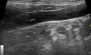
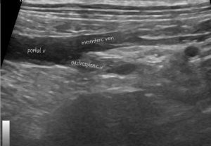
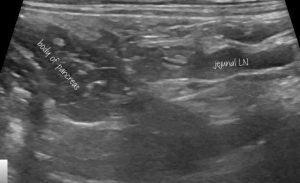
This is what you are seeing, in sequence from dorsal to ventral;
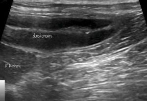
The duodenum is consistently the most dorsal and superficial loop of small intestine in the right cranial abdomen in smaller dogs. In larger dogs it sometimes lies inside the caecum or even jejunum. Either way it’s the ‘fattest’ loop of small bowel and has the thickest mucosa. The kidney is an obvious reference point.
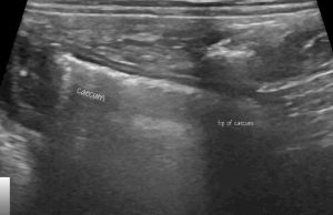
The caecum is curved: in this clip you first see the gas-filled distal part. As the video pans ventrally you can follow round the caecum into the ICCJ. You may have to slide the probe contact point slightly caudal to centre over the junction.
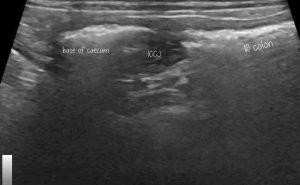
…and there it is. In dogs, the ileum enters the ICCJ on its axial aspect and from caudally.
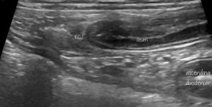
If the caecum and right colon have more gas/faeces in the lumen then the shadow from that may obscure the ileum and ICCJ. You will see the abaxial wall of the caecum and right colon and below that hyperechoic faeces. At the junction between the two will be a dip or notch. If you apply gentle but firm pressure here for a while then the gut content will normally disperse such that you can see the ileum and ICCJ beneath the notch.
Cats are a bit different…..


